Journal of Jilin University(Medicine Edition) ›› 2022, Vol. 48 ›› Issue (6): 1593-1598.doi: 10.13481/j.1671-587X.20220627
• Clinical medicine • Previous Articles Next Articles
Renal solitary fibrous tumor complicated with hydronephrosis: A case report and literature review
Pengxiang HUI,Xiao YANG,Xu WANG,Ming ZHANG,Haitao FAN,Huikang YU,Yinchun WANG,Qun ZHAO,Gaowen TANG,Ranwei LI( )
)
- Department of Urology,Second Hospital,Jilin University,Changchun 130041,China
-
Received:2021-12-15Online:2022-11-28Published:2022-12-07 -
Contact:Ranwei LI E-mail:ranwei1968@sina.com
CLC Number:
- R737.11
Cite this article
Pengxiang HUI,Xiao YANG,Xu WANG,Ming ZHANG,Haitao FAN,Huikang YU,Yinchun WANG,Qun ZHAO,Gaowen TANG,Ranwei LI. Renal solitary fibrous tumor complicated with hydronephrosis: A case report and literature review[J].Journal of Jilin University(Medicine Edition), 2022, 48(6): 1593-1598.
share this article
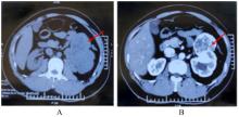
Fig. 1
CT images of patient with renal SFT complicated with hydronephrosis"

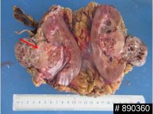
Fig. 2
General morphology of left renal tumor(Bar=1.0 cm)"

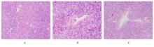
Fig. 3
Pathomorphology of tumor tissue of patient with renal SFT complicated with hydronephrosis observed by HE staining"
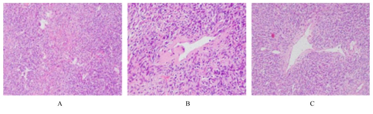
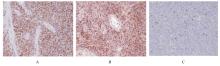
Fig. 4
Results of immunohistochemical staining of tumor tissue of patient with renal SFT complicated with hydronephrosis (×100)"


Fig. 5
Postoperative CT image of patient with renal SFT complicated with hydronephrosis"

| 1 | KLEMPERER P, COLEMAN B R. Primary neoplasms of the pleura. A report of five cases [J]. Am J Ind Med, 1992, 22(1): 1-31. |
| 2 | STOUT A P, MURRAY M R. Hemangiopericytoma: a vascular tumor featuring zimmermann’s pericytes[J]. Ann Surg, 1942, 116(1): 26-33. |
| 3 | DONG B, ZHANG J J, WANG G, et al. Renal solitary fibrous tumour: a rare pathological entity[J]. Can Urol Assoc J, 2014, 8(9/10): E657-E659. |
| 4 | 王晓龙, 陈鸿远, 王朝明, 等. 3例前列腺孤立性纤维性肿瘤报道及文献复习[J].现代肿瘤医学,2021,29(19): 3468-3470. |
| 5 | GELB A B, SIMMONS M L, WEIDNER N. Solitary fibrous tumor involving the renal capsule[J]. Am J Surg Pathol, 1996, 20(10): 1288-1295. |
| 6 | 张建兵, 金 梅, 朱 涛, 等. 肾孤立性纤维性肿瘤一例[J]. 中华病理学杂志, 2014, 43(1): 44-45. |
| 7 | MARTIN-BROTO J, MONDAZA-HERNANDEZ J L, MOURA D S, et al. A comprehensive review on solitary fibrous tumor: new insights for new horizons[J]. Cancers, 2021, 13(12): 2913. |
| 8 | 赵大华, 吴淑华, 朱玉红, 等. 肾恶性孤立性纤维性肿瘤一例报告[J]. 中华泌尿外科杂志, 2008, 29(3): 195. |
| 9 | 张玉华, 高子芬, 董格红, 等. 孤立性纤维性肿瘤7例临床病理分析[J]. 诊断病理学杂志, 2020, 27(11): 816-821. |
| 10 | HAN Y N, ZHANG Q F, YU X M, et al. Immunohistochemical detection of STAT6, CD34, CD99 and BCL-2 for diagnosing solitary fibrous tumors/hemangiopericytomas[J]. Int J Clin Exp Pathol, 2015, 8(10): 13166-13175. |
| 11 | SCOTLANDI K, ZUNTINI M, MANARA M C,et al. CD99 isoforms dictate opposite functions in tumour malignancy and metastases by activating or repressing c-Src kinase activity[J]. Oncogene, 2007, 26(46): 6604-6618. |
| 12 | CHMIELECKI J, CRAGO A M, ROSENBERG M, et al. Whole-exome sequencing identifies a recurrent NAB2-STAT6 fusion in solitary fibrous tumors[J]. Nat Genet, 2013, 45(2): 131-132. |
| 13 | BARTHELMEβ S, GEDDERT H, BOLTZE C, et al. Solitary fibrous tumors/hemangiopericytomas with different variants of the NAB2-STAT6 gene fusion are characterized by specific histomorphology and distinct clinicopathological features[J]. Am J Pathol, 2014, 184(4): 1209-1218. |
| 14 | 丁志燕, 王艳芬, 王 璇, 等. 信号转导及转录激活因子6在孤立性纤维性肿瘤中的表达和意义[J]. 中华病理学杂志, 2017, 46(4): 235-239. |
| 15 | YAZAKI T, SATOH S, IIZUMI T, et al. Solitary fibrous tumor of renal pelvis[J]. Int J Urol, 2001, 8(9): 504-508. |
| 16 | SASAKI H, KURIHARA T, KATSUOKA Y, et al. Distant metastasis from benign solitary fibrous tumor of the kidney[J]. Case Rep Nephrol Urol, 2013,3(1): 1-8. |
| 17 | BEZERRA E S, ANDRIGHETTO O P, COSTA M V S,et al. Bilateral renal involvement by solitary fibrous tumor - Report of a case in the post-WHO/2016 era[J]. Urol Case Rep, 2018, 17: 7-9. |
| 18 | O’NEILL A C, TIRUMANI S H, DO W S, et al. Metastatic patterns of solitary fibrous tumors: a single-institution experience[J]. AJR Am J Roentgenol, 2017, 208(1): 2-9. |
| 19 | DEMICCO E G, PARK M S, ARAUJO D M, et al. Solitary fibrous tumor: a clinicopathological study of 110 cases and proposed risk assessment model[J]. Mod Pathol, 2012, 25(9): 1298-1306. |
| 20 | BALDI G G, STACCHIOTTI S, MAURO V, et al. Solitary fibrous tumor of all sites: outcome of late recurrences in 14 patients[J]. Clin Sarcoma Res, 2013, 3: 4. |
| 21 | DEMICCO E G, WAGNER M J, MAKI R G, et al. Risk assessment in solitary fibrous tumors: validation and refinement of a risk stratification model[J]. Mod Pathol, 2017, 30(10): 1433-1442. |
| 22 | SALAS S, RESSEGUIER N, BLAY J Y, et al. Prediction of local and metastatic recurrence in solitary fibrous tumor: construction of a risk calculator in a multicenter cohort from the French Sarcoma Group (FSG) database[J]. Ann Oncol, 2017, 28(8): 1979-1987. |
|