Journal of Jilin University(Medicine Edition) ›› 2026, Vol. 52 ›› Issue (2): 330-339.doi: 10.13481/j.1671-587X.20260204
• Research in basic medicine • Previous Articles Next Articles
Improvement effect of fingolimod on hepatic fibrosis in type 2 diabetes mellitus mice and its mechanism
Shu LI1,2,Jiaqi GUO3,Wansong LI4,Yanfeng ZHEN2,Hongjia ZHAI4,Jie LI4,Hui FANG2( )
)
- 1.Department of Internal Medicine,School of Clinical Medicine,North China University of Science and Technology,Tangshan 063000,China
2.Department of Endocrinology,Tangshan Workers’ Hospital,Tangshan 063000,China
3.Department of Critical Care Medicine,Central Hospital,Longhua District,Guangdong Province,Shenzhen 518000,China
4.Department of Internal Medicine,School of Clinical Medicine,Hebei Medical University,Shijiazhuang 050000,China
-
Received:2025-05-14Accepted:2025-07-06Online:2026-03-28Published:2026-04-15 -
Contact:Hui FANG E-mail:fanghui2818@126.com
CLC Number:
- R587.2
Cite this article
Shu LI,Jiaqi GUO,Wansong LI,Yanfeng ZHEN,Hongjia ZHAI,Jie LI,Hui FANG. Improvement effect of fingolimod on hepatic fibrosis in type 2 diabetes mellitus mice and its mechanism[J].Journal of Jilin University(Medicine Edition), 2026, 52(2): 330-339.
share this article
Tab. 1
Primer sequences of RT-qPCR"
| Gene | Forward sequence(5'-3') | Reverse sequence(5'-3') |
|---|---|---|
| β-actin | GTGACGTTGACATCCGTAAAGA | GCCGGACTCATCGTACTCC |
| JAK1 | AGTGCAGTATCTCTCCTCTCTG | GATTCGGTTCGGAGCGTACC |
| JAK2 | GGAATGGCCTGCCTTACAATG | TGGCTCTATCTGCTTCACACAAT |
| STAT1 | TCACAGTGGTTCGAGCTTCAG | CGAGACATCATAGGCAGCGTG |
| STAT3 | CACTTGGATTGAGAGTCAAGAC | AGGAATCGGCTATATTGCTGGT |
| IFN-γ | GCCACGGCACAGTCATTGA | TGCTGATGGCCTGATTGTCTT |
| IL-6 | CTGCAAGAGACTTCCATCCAG | AGTGGTATAGACAGGTCTGTTGG |
| α-SMA | TCGGATACTTCAGCGTCA | GGGAGTAATGGTTGGAATG |
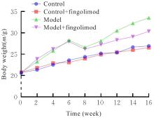
Fig. 1
Body weights of mice in various groups"
Tab.2
Liver coefficients and FBG levels of mice in various groups"
| Group | Liver coefficient(η/%) | FBG level[cB/(mmol·L-1)] |
|---|---|---|
| Control | 3.54±0.18 | 5.80±1.06 |
| Control+fingolimod | 3.65±0.29 | 5.31±0.95 |
| Model | 4.69±0.31* | 21.90±1.96* |
| Model+fingolimod | 4.17±0.24△ | 15.68±1.65△ |
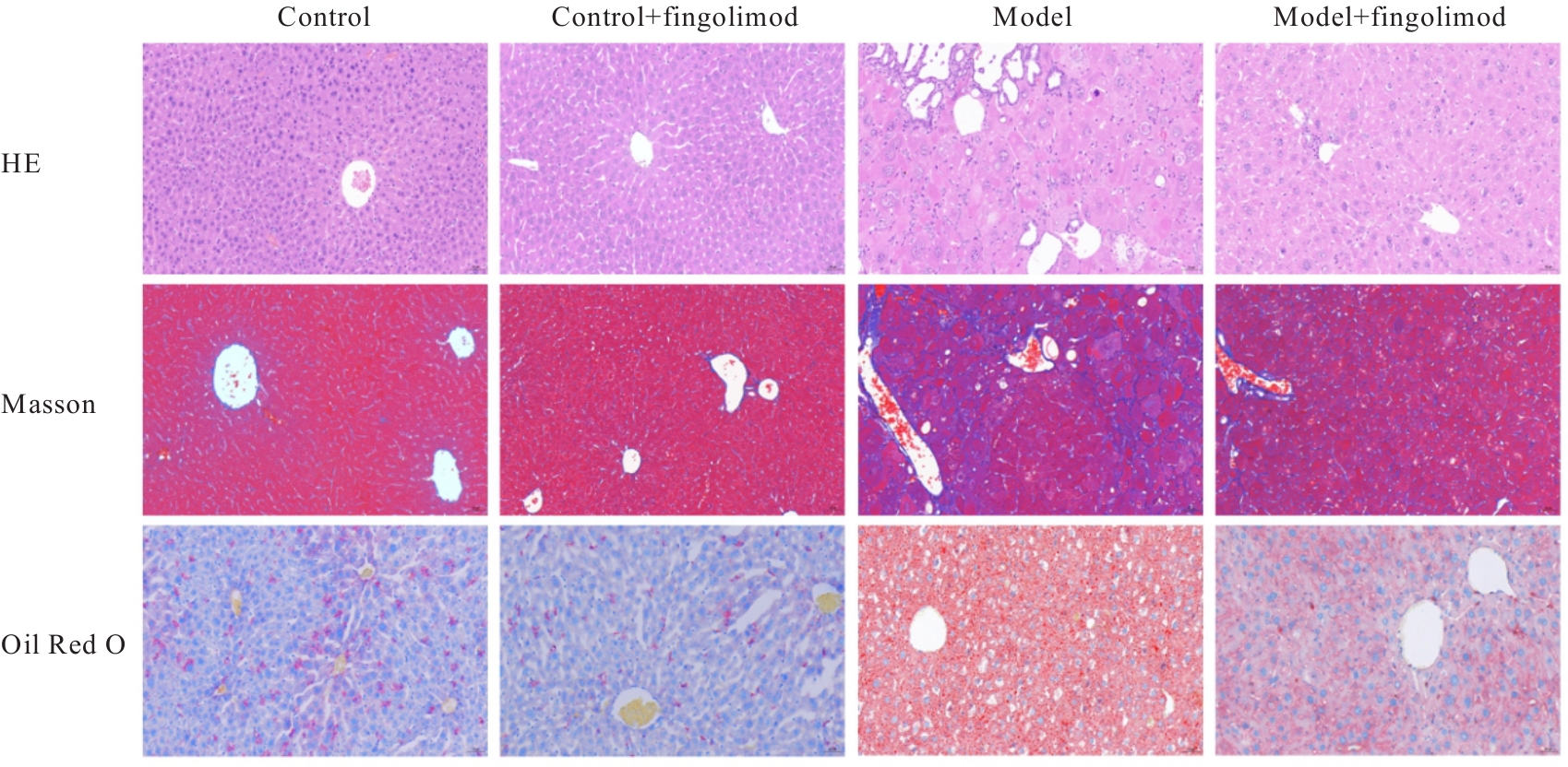
Fig. 2
Pathomorphology of liver tissue of mice in various groups (×200)"
Tab. 3
Hepatic CVF and lipid droplet area proportions of mice in various groups"
| Group | CVF | Lipid droplet area ratio proportion |
|---|---|---|
| Control | 2.23±2.37 | 0.46±1.13 |
| Control+fingolimod | 3.14±2.08 | 0.07±1.85 |
| Model | 38.61±2.95* | 44.50±1.47* |
| Model+fingolimod | 22.15±2.07△ | 10.17±1.43△ |
Tab.4
Activities of ALT and AST, and levels of TC, TG, LDL-C, and HDL-C in serum of mice in various groups"
| Group | ALT[λB/(U·L-1)] | AST[λB/(U·L-1)] | TC[cB/(mmol·L-1)] | TG[cB/(mmol·L-1)] | LDL-C[cB/(mmol·L-1)] | HDL-C[cB/(mmol·L-1)] |
|---|---|---|---|---|---|---|
| Control | 24.74±1.933 | 29.89±2.581 | 1.51±0.100 | 0.65±0.061 | 0.40±0.036 | 1.41±0.031 |
| Control+fingolimod | 23.63±2.276 | 30.24±2.839 | 1.54±0.136 | 0.53±0.067 | 0.40±0.044 | 1.40±0.062 |
| Model | 74.13±8.297* | 90.55±6.269* | 2.45±0.133* | 2.10±0.188* | 1.28±0.074* | 0.71±0.053* |
| Model+fingolimod | 36.73±4.480△ | 44.37±3.360△ | 2.02±0.0540△ | 1.20±0.119△ | 0.71±0.049△ | 1.02±0.093△ |
Tab.5
Expression levels of JAK1, JAK2, STAT1, STAT3, IFN-γ, IL-6, and α-SMA mRNA in liver tissue of mice in various groups"
| Group | JAK1 | JAK2 | STAT1 | STAT3 | IFN-γ | IL-6 | α-SMA |
|---|---|---|---|---|---|---|---|
| Control | 1.00±0.08 | 1.00±0.15 | 1.00±0.06 | 1.00±0.10 | 1.00±0.08 | 1.00±0.13 | 1.02±0.23 |
| Control+fingolimod | 1.15±0.19 | 0.93±0.08 | 0.94±0.16 | 0.91±0.12 | 0.93±0.13 | 1.07±0.19 | 0.69±0.13 |
| Model | 5.39±0.76* | 6.01±0.48* | 0.15±0.03* | 3.92±0.22* | 0.03±0.01* | 2.99±0.15* | 8.15±0.28* |
| Model+fingolimod | 2.78±0.49△ | 3.00±0.49△ | 0.48±0.06△ | 2.84±0.20△ | 0.27±0.03△ | 1.56±0.11△ | 2.45±0.42△ |
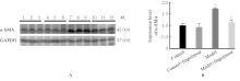
Fig. 3
Electrophoregram(A) and histogram(B) of expressions of α-SMA protein in liver tissue of mice in various groups"
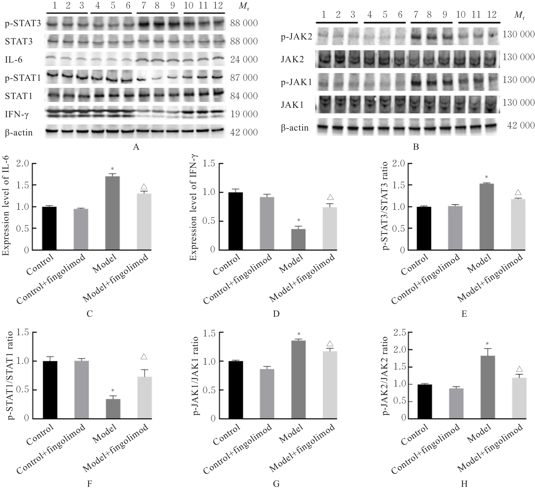
Fig. 4
Electrophoregrams(A,B) and histograms(C-H) of expressions of IL-6, IFN-γ, p-STAT1, STAT1, p-STAT3, STAT3, p-JAK1, JAK1, p-JAK2 and JAK2 proteins in liver tissue of mice in various groups"
| [1] | YAMAMURA S, ESLAM M, KAWAGUCHI T, et al. MAFLD identifies patients with significant hepatic fibrosis better than NAFLD[J]. Liver Int, 2020, 40(12): 3018-3030. |
| [2] | ZHAO J, LIU L, CAO Y Y, et al. MAFLD as part of systemic metabolic dysregulation[J]. Hepatol Int, 2024, 18(): 834-847. |
| [3] | WANG Q X, LU T M, SONG P, et al. Glycyrrhizic acid ameliorates hepatic fibrosis by inhibiting oxidative stress via AKR7A2[J]. Phytomedicine, 2024, 133: 155878. |
| [4] | CHEN J Z, GE J M, CHEN W S, et al. UPLC-Q-TOF-MS based investigation into the bioactive compounds and molecular mechanisms of Lamiophlomis Herba against hepatic fibrosis[J]. Phytomedicine, 2023, 121: 155085. |
| [5] | GUO M M, WANG Z D, DAI J Y, et al. Glycyrrhizic acid alleviates liver fibrosis in vitro and in vivo via activating CUGBP1-mediated IFN-γ/STAT1/Smad7 pathway[J]. Phytomedicine, 2023, 112: 154587. |
| [6] | HIGASHI T, FRIEDMAN S L, HOSHIDA Y. Hepatic stellate cells as key target in liver fibrosis[J]. Adv Drug Deliv Rev, 2017, 121: 27-42. |
| [7] | 韩 超, 王丽娟, 王鹏源, 等. IL-6/JAK/STAT3信号通路在纤维化疾病中作用的研究进展[J]. 中国病理生理杂志, 2022, 38(12): 2285-2290. |
| [8] | WANG Z F, KAWABORI M, HOUKIN K. FTY720 (fingolimod) ameliorates brain injury through multiple mechanisms and is a strong candidate for stroke treatment[J]. Curr Med Chem, 2020, 27(18): 2979-2993. |
| [9] | ROHRBACH T D, ASGHARPOUR A, MACZIS M A, et al. FTY720/fingolimod decreases hepatic steatosis and expression of fatty acid synthase in diet-induced nonalcoholic fatty liver disease in mice[J]. J Lipid Res, 2019, 60(7): 1311-1322. |
| [10] | YE H J, SUN M Y, JIN Z J, et al. FTY-720 alleviates diabetes-induced liver injury by inhibiting oxidative stress and inflammation[J]. Fundam Clin Pharmacol, 2023, 37(5): 960-970. |
| [11] | LOU D, FANG Q, HE Y H, et al. Oxymatrine alleviates high-fat diet/streptozotocin-induced non-alcoholic fatty liver disease in C57BL/6 J mice by modulating oxidative stress, inflammation and fibrosis[J]. Biomed Pharmacother, 2024, 174: 116491. |
| [12] | SOOD A, FERNANDES V, PREETI K, et al. Sphingosine 1 phosphate lyase inhibition rescues cognition in diabetic mice by promoting anti-inflammatory microglia[J]. Behav Brain Res, 2023, 446: 114415. |
| [13] | SOOD A, FERNANDES V, PREETI K, et al. Fingolimod alleviates cognitive deficit in type 2 diabetes by promoting microglial M2 polarization via the pSTAT3-jmjd3 axis[J]. Mol Neurobiol, 2023, 60(2): 901-922. |
| [14] | YIN Z Y, FAN L N, WEI L P, et al. FTY720 protects cardiac microvessels of diabetes: a critical role of S1P1/3 in diabetic heart disease[J]. PLoS One, 2012, 7(8): e42900. |
| [15] | LIU Z L, XU B G, DING Y P, et al. Guizhi Fuling pill attenuates liver fibrosis in vitro and in vivo via inhibiting TGF-β1/Smad2/3 and activating IFN-γ/Smad7 signaling pathways[J]. Bioengineered, 2022, 13(4): 9357-9368. |
| [16] | WENG H L, MERTENS P R, GRESSNER A M, et al. IFN-gamma abrogates profibrogenic TGF-beta signaling in liver by targeting expression of inhibitory and receptor Smads[J]. J Hepatol, 2007, 46(2): 295-303. |
| [17] | ZAI W J, CHEN W, LUAN J Y, et al. Dihydroquercetin ameliorated acetaminophen-induced hepatic cytotoxicity via activating JAK2/STAT3 pathway and autophagy[J]. Appl Microbiol Biotechnol, 2018, 102(3): 1443-1453. |
| [18] | YU H, LEE H, HERRMANN A, et al. Revisiting STAT3 signalling in cancer: new and unexpected biological functions[J]. Nat Rev Cancer, 2014, 14(11): 736-746. |
| [19] | GARRIS C S, WU L F, ACHARYA S, et al. Defective sphingosine 1-phosphate receptor 1 (S1P1) phosphorylation exacerbates TH17-mediated autoimmune neuroinflammation[J]. Nat Immunol, 2013, 14(11): 1166-1172. |
| [20] | LIU Y, DENG J H, WANG L, et al. S1PR1 is an effective target to block STAT3 signaling in activated B cell-like diffuse large B-cell lymphoma[J]. Blood, 2012, 120(7): 1458-1465. |
| [21] | GU Y J, SUN W Y, ZHANG S, et al. Targeted blockade of JAK/STAT3 signaling inhibits proliferation, migration and collagen production as well as inducing the apoptosis of hepatic stellate cells[J]. Int J Mol Med, 2016, 38(3): 903-911. |
| [22] | ZHAO J J, JIAO Y, SONG Y P, et al. Stanniocalcin 2 ameliorates hepatosteatosis through activation of STAT3 signaling[J]. Front Physiol, 2018, 9: 873. |
| [1] | Xiaoqian TANG,Shengcong WEN,Zhenya DONG,Jingyi CHEN,Yu CAO,Yunhua ZHANG. Improvement effect of engineered exosomes delivering ANGPTL6 mRNA on liver fibrosis in mice [J]. Journal of Jilin University(Medicine Edition), 2025, 51(6): 1452-1463. |
| [2] | Huixian XU,Hui XU,Jishu QUAN,Feng ZHENG. Inhibitory effect of iridoid glycosides from Boschniakia rossica on hepatic preneolasia of rats and its mechanism [J]. Journal of Jilin University(Medicine Edition), 2025, 51(4): 887-895. |
| [3] | Yanbin ZHANG,Guangye GUO,Caihua ZHENG,Xinyan LIU. Research progress in association between Helicobacter pylori and metabolic syndrome and its effect on occurrence and development of metabolic syndrome [J]. Journal of Jilin University(Medicine Edition), 2024, 50(6): 1757-1762. |
| [4] | Yajie GE,Wen XU,Shimin GUAN,Lina WANG. Research progress in etiology and pathogenesis of polycystic ovary syndrome [J]. Journal of Jilin University(Medicine Edition), 2024, 50(1): 288-294. |
| [5] | Jiaxin WANG,Zhenqi WANG,Xuan ZHANG. Expressions of CDKAL1 gene and its splice isomers in peripheral blood lymphocytes of patients with type 2 diabetes mellitus and their clinical significances [J]. Journal of Jilin University(Medicine Edition), 2023, 49(5): 1290-1295. |
| [6] | Jing GUAN,Shen HA,Hao YUAN,Ying CHEN,Pengju LIU,Zhi LIU,Shuang JIANG. Protective effect of Modified Xiao-Xian-Xiong Decoction on liver injury in rats with type 2 diabete mellitus and its mechanism [J]. Journal of Jilin University(Medicine Edition), 2023, 49(3): 608-616. |
| [7] | Xiaowei HUANG,Siqi ZHANG,Yixin ZHANG,Bo LIU,Xin WANG,Fenglan JI,Runze YANG,Huibo XU,Tao DING. Relationship between syndrome manifestations and differentially expressed genes in rat model of type 2 diabetes mellitus with Qi and Yin deficiency explored through transcriptomics [J]. Journal of Jilin University(Medicine Edition), 2023, 49(3): 625-633. |
| [8] | Lingyao XU,Shutang WEI,Yong DONG,Zhenglu SUN,Junbo ZHAO,Dazheng HAN. Regulatory effect of lncRNA MALAT1 on activation of hepatic stellate cell and its mechanism [J]. Journal of Jilin University(Medicine Edition), 2023, 49(3): 697-705. |
| [9] | Junxiong ZHAO,Qian WU,Liangui NIE,Shengquan LIU,Zhentao JIANG,Jian CHEN,Ting XIAO,Jun YANG. Ameliorative effect of SO2 on myocardial fibrosis in type 2 diabetes mellitus rats and its mechanism [J]. Journal of Jilin University(Medicine Edition), 2023, 49(1): 8-14. |
| [10] | Yihua LI,Tao WEN,Yongri QIAN,Chunshan ZHAO. Correlation analysis on relationships between levels of serum uric acid and γ-glutamyl transpeptidase and metabolic syndrome in healthy physical examination population [J]. Journal of Jilin University(Medicine Edition), 2022, 48(6): 1605-1613. |
| [11] | Mingliu GU,Fengyuan LIN,Xuefeng WU,Jianing ZHOU,Xuechun LU,Peige DU,Liping AN. Improvement effect of Phellinus igniarius acidic polysaccharide on liver fibrosis induced by bile duct ligation in mice and its mechanism [J]. Journal of Jilin University(Medicine Edition), 2022, 48(5): 1167-1174. |
| [12] | Yongxin ZHAI,Huaifeng TA,Yi ZHANG,Qijia SUN,Ming ZHANG,Shuqiang FENG,Ranwei LI. Effect of preoperative glycemic level on infection-related complications of diabetic patients after flexible ureteroscopic lithotripsy [J]. Journal of Jilin University(Medicine Edition), 2022, 48(3): 766-772. |
| [13] | Yang ZHENG,Jiahui WANG,Yue PENG,Xianling YUAN,Lei WANG,Tiejian ZHAO. Influence of curcumol in structure of liver sinusoidal endothelial cells of mice and its inhibitiory effect on intrahepatic angiogenesis [J]. Journal of Jilin University(Medicine Edition), 2021, 47(5): 1124-1130. |
| [14] | Zhouguang WU,Bin WANG,Zhen CHENG,Wenjie ZHANG,Taoyan ZUO,Siqi CHEN,Jingru FU. Analysis on correlation of GPC3 and α-SMA expressions with liver function-related indexes in liver tissue of children with biliary atresia [J]. Journal of Jilin University(Medicine Edition), 2021, 47(5): 1237-1243. |
| [15] | Rongjun JIA,Liman MA,Lihua LI. Protective effect of calcitriol on hepatic fibrosis induced by bile duct ligation in mice and its mechanism [J]. Journal of Jilin University(Medicine Edition), 2021, 47(2): 257-264. |
|