Journal of Jilin University(Medicine Edition) ›› 2025, Vol. 51 ›› Issue (3): 703-715.doi: 10.13481/j.1671-587X.20250315
• Research in clinical medicine • Previous Articles
Bioinformatics analysis on adjustment effect of colorectal liver metastases model in mice based on complement alternative pathway and its experimental verification
Changyu SHI1,Yong LI1,Jing DENG1,Chunmei PIAO2,Ming JIN1( )
)
- 1.Department of Biochemistry and Molecular Biology,School of Medical Sciences,Yanbian University,Yanji 133000,China
2.Affiliated Beijing Anzhen Hospital,Capital Medical University,Beijing Institute of Heart Lung and Blood Vessel Diseases,Beijing 100029,China
-
Received:2024-05-09Accepted:2024-08-06Online:2025-05-28Published:2025-07-18 -
Contact:Ming JIN E-mail:jinming@ybu.edu.cn
CLC Number:
- R735.3
Cite this article
Changyu SHI,Yong LI,Jing DENG,Chunmei PIAO,Ming JIN. Bioinformatics analysis on adjustment effect of colorectal liver metastases model in mice based on complement alternative pathway and its experimental verification[J].Journal of Jilin University(Medicine Edition), 2025, 51(3): 703-715.
share this article
Tab.1
Sequences of primers"
| Primer | Forward(5'-3') | Reverse(5'-3') |
|---|---|---|
| C1q | GAAGGGCGTGAAAGGCAATC | CAAGCGTCATTGGGTTCTGC |
| C2 | AGAGTGCCGAACTCATGGTG | GGCTGAGAGGCAAAGGTGAT |
| C3 | ATAAAGAGCCAGCGGCTACA | GAATGAAGGGGTAAGGGGCA |
| C3aR | CCCCAAGACATTGCCTCCAT | GACTGTGTTCACGGTCGTCT |
| C5 | TGGTTCCTTCAGCACGACTC | CAGACTGCGTCAGCCACTAA |
| C5aR | TCCTTCAGAAGAGTTGCCTGC | TTCTGTGGTAACCAGCGACG |
| DAF | GCTCAATTAACTGCGGCTCAA | ACAGAGACAGCGACAGCAAC |
| FB | ATGGGGACAAGAAAGCCAGTT | TAGCCTTGGGCCTTTGTAGC |
| FD | TCGAAGGTGTGGTTACGTGG | TCGATCCACATCCGGTAGGA |
| GADPH | AATGCATCCTGCACCACC | ATGCCAGTGAGCTTCCCG |
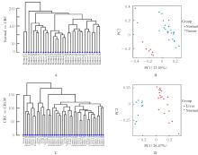
Fig. 1
Cluster analysis and PCA analysis on normal colon tissue samples and CRC tissue samples and CRC tissue samples and CRC liver metastasis tissue samples"
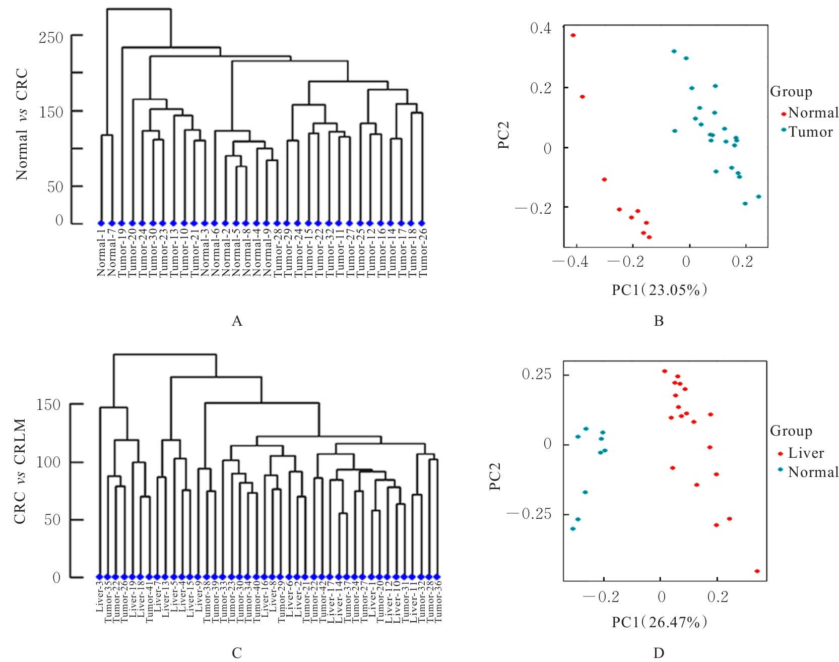
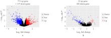
Fig. 2
Volcanic maps of normal colon tissue samples and CRC tissue samples and CRC tissue samples and CRC liver metastasis tissue samples"

Fig. 3
DEGs heat maps of normal colon tissue samples and CRC tissue samples and CRC tissue samples and CRC liver metastasis liver tissue samples"
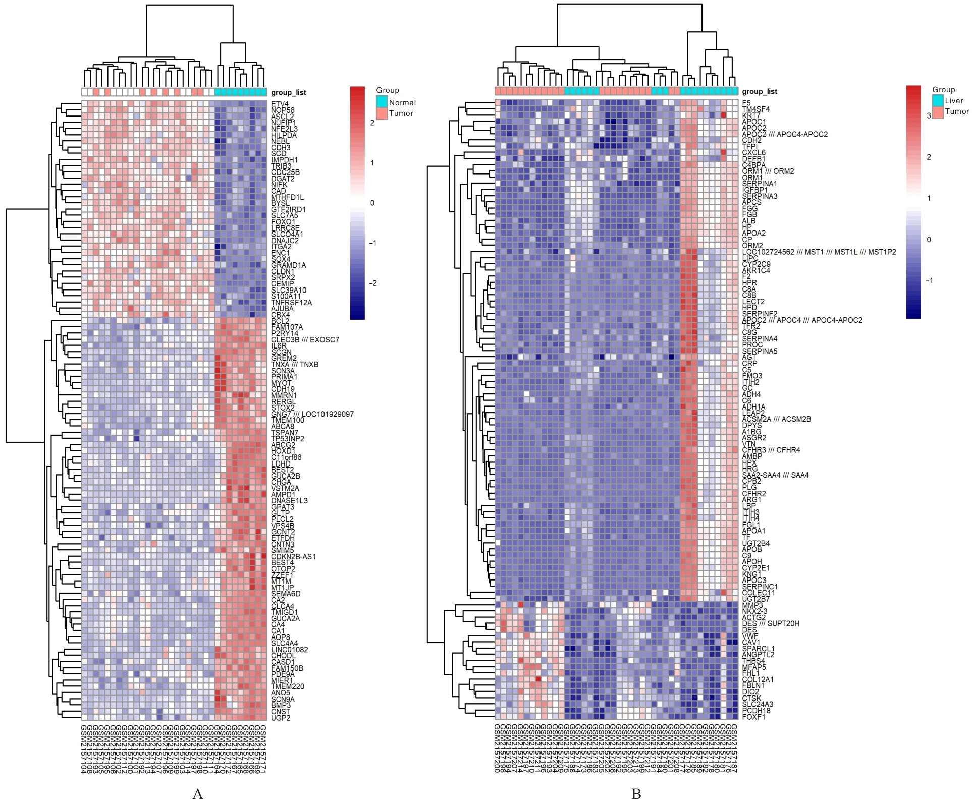
Fig. 4
GO functional enrichment maps of DGEs of normal colon tissue samples and CRC samples and CRC tissue samples and CRC liver metastasis tissue samples"

Fig. 5
KEGG enrichment map of DGEs of normal colon tissue samples and CRC tissue samples and CRC tissue samples and CRC liver metastasis tissue samples"
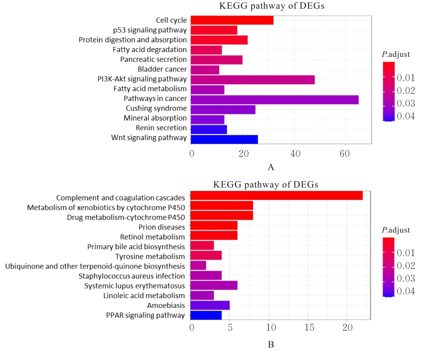
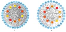
Fig. 6
PPI network diagrams of DGEs of normal colon tissue sample and CRC samples and CRC and CRC liver metastasis tissue samples"
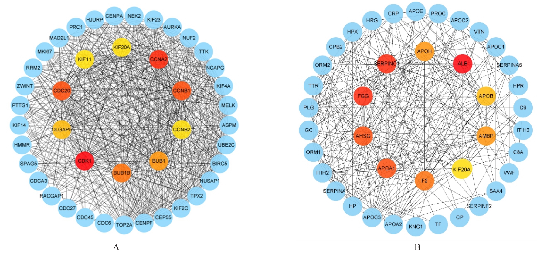
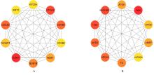
Fig. 7
Top 10 Hub genes of normal colon tissue samples and CRC tissue samples and CRC tissue samples and CRC liver metastasis tissue samples"
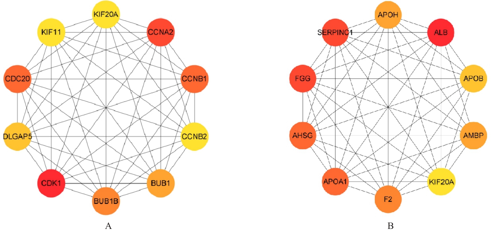
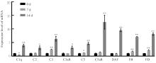
Fig. 8
Expression levels of complement pathway-related genes in CRC liver metastasis liver tissue of mice in various groups detected by RT-qPCR method"
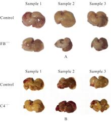
Fig. 9
General morphology of liver of mice after knockout of FB and C4 factors"

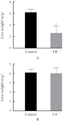
Fig. 10
Liver weights of mice after knockout of FB and C4 factors"


Fig. 11
Infiltration of CRC liver metastases of mice in two groups after knockout of FB observed by HE staining"
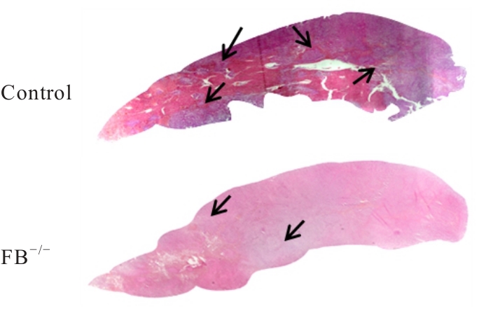
Fig. 12
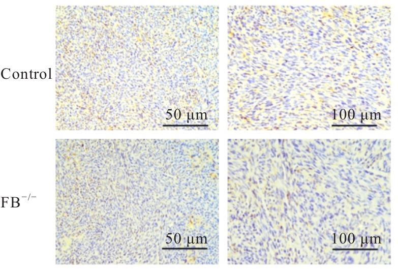
Macrophage infiltration in CRC liver metastases of mice in two groups after knockout of FB observed by immunohistochemisty staining"
| [1] | BENSON A B, VENOOK A P, AL-HAWARY M M, et al. Colon cancer, version 2.2021, NCCN clinical practice guidelines in oncology[J]. J Natl Compr Canc Netw, 2021, 19(3): 329-359. |
| [2] | TSAI M S, SU Y H, HO M C, et al. Clinicopathological features and prognosis in resectable synchronous and metachronous colorectal liver metastasis[J]. Ann Surg Oncol, 2007, 14(2): 786-794. |
| [3] | MAURI G, BONAZZINA E, AMATU A, et al. The evolutionary landscape of treatment for BRAFV600E mutant metastatic colorectal cancer[J]. Cancers (Basel), 2021, 13(1): 137. |
| [4] | RUFFINELLI J C, SANTOS VIVAS C, SANZ-PAMPLONA R, et al. New advances in the clinical management of RAS and BRAF mutant colorectal cancer patients[J]. Expert Rev Gastroenterol Hepatol, 2021, 15(1): 65-79. |
| [5] | LUO Q X, CHEN D K, FAN X J, et al. KRAS and PIK3CA bi-mutations predict a poor prognosis in colorectal cancer patients: a single-site report[J]. Transl Oncol, 2020, 13(12): 100874. |
| [6] | WANG D W, SU F, YANG L J, et al. Bioinformatics analysis and identification of potential genes associated with pathogenesis and prognosis of gastric cancer[J]. Curr Med Sci, 2022, 42(2): 357-372. |
| [7] | TALAAT I M, ELEMAM N M, SABER-AYAD M. Complement system: an immunotherapy target in colorectal cancer[J]. Front Immunol, 2022, 13: 810993. |
| [8] | CHANG H L, JIN L, XIE P Y, et al. Complement C5 is a novel biomarker for liver metastasis of colorectal cancer[J]. J Gastrointest Oncol, 2022, 13(5): 2351-2365. |
| [9] | PIAO C M, CAI L, QIU S L, et al. Complement 5a enhances hepatic metastases of colon cancer via monocyte chemoattractant protein-1-mediated inflammatory cell infiltration[J]. J Biol Chem, 2015, 290(17): 10667-10676. |
| [10] | IRIZARRY R A, HOBBS B, COLLIN F, et al. Exploration, normalization, and summaries of high density oligonucleotide array probe level data[J]. Biostatistics, 2003, 4(2): 249-264. |
| [11] | WEI R Q, ZHANG W M, LIANG Z, et al. Identification of signal pathways and hub genes of pulmonary arterial hypertension by bioinformatic analysis[J]. Can Respir J, 2022, 2022: 1394088. |
| [12] | RITCHIE M E, PHIPSON B, WU D, et al. Limma powers differential expression analyses for RNA-sequencing and microarray studies[J]. Nucleic Acids Res, 2015, 43(7): e47. |
| [13] | YOUNG A, WHITEHOUSE N, CHO J, et al. OntologyTraverser: an R package for GO analysis[J]. Bioinformatics, 2005, 21(2): 275-276. |
| [14] | KANEHISA M. The KEGG database[J]. Novartis Found Symp, 2002, 247: 91-101; discussion101-103, 119-128, 244-252. |
| [15] | 崔 巍, 杜 杰, 杨 敏, 等. 小鼠结直肠癌肝转移模型的建立及其炎症微环境分析[J]. 中国癌症杂志, 2012, 22(8): 566-572, 582. |
| [16] | SCHILLING J D, MACHKOVECH H M, KIM A H J, et al. Macrophages modulate cardiac function in lipotoxic cardiomyopathy[J]. Am J Physiol Heart Circ Physiol, 2012, 303(11): H1366-H1373. |
| [17] | HAN Y H, MUN J G, JEON H D, et al. Betulin inhibits lung metastasis by inducing cell cycle arrest, autophagy, and apoptosis of metastatic colorectal cancer cells[J]. Nutrients, 2019, 12(1): 66. |
| [18] | MAREI H E, ALTHANI A, AFIFI N, et al. p53 signaling in cancer progression and therapy[J]. Cancer Cell Int, 2021, 21(1): 703. |
| [19] | ADMASU F T, DEJENIE T A, AYEHU G W, et al. Evaluation of thromboembolic event, basic coagulation parameters, and associated factors in patients with colorectal cancer: a multicenter study[J]. Front Oncol, 2023, 13: 1143122. |
| [20] | KROONE C, TIEKEN C, KOCATÜRK B, et al. Tumor-expressed factor Ⅶ is associated with survival and regulates tumor progression in breast cancer[J]. Blood Adv, 2023, 7(11): 2388-2400. |
| [21] | DAHA M R, SEELEN M. Novel approaches to control of the alternative complement pathway for the treatment of C3 glomerulopathies[J]. J Am Soc Nephrol, 2018, 29(8): 2032-2033. |
| [22] | COSS S L, ZHOU D L, CHUA G T, et al. The complement system and human autoimmune diseases[J]. J Autoimmun, 2023, 137: 102979. |
| [23] | AYKUT B, PUSHALKAR S, CHEN R N, et al. The fungal mycobiome promotes pancreatic oncogenesis via activation of MBL[J]. Nature, 2019, 574(7777): 264-267. |
| [24] | 郭楠楠, 王兴智. 补体B因子的研究进展[J]. 国际免疫学杂志, 2022, 45(1): 6. |
| [25] | 路 平, 魏少忠, 梁新军. 补体系统与肿瘤免疫的研究进展[J]. 中国肿瘤临床, 2021, 48(13): 681-685. |
| [26] | 陈立颖, 朱 莹, 封 奕, 等. 结肠癌组织中补体C5a/C5aR通路活化上调FGL2的表达[J]. 第三军医大学学报, 2016, 38(6): 575-579. |
| [27] | WU P, SHI J Y, SUN W, et al. The prognostic value of plasma complement factor B (CFB) in thyroid carcinoma[J]. Bioengineered, 2021, 12(2): 12854-12866. |
| [28] | MANTOVANI A, ALLAVENA P, MARCHESI F, et al. Macrophages as tools and targets in cancer therapy[J]. Nat Rev Drug Discov, 2022, 21(11): 799-820. |
| [1] | Wenchang CAI,Yuqi LIU,Han WANG,Helin WANG,Zhenjiang WANG,Zishen XIAO,Shiyuan MA,Liping AN,Yanbo LIU. Expression of protein kinase D2 in bladder cancer tissue and its effect on tumor immune microenvironment [J]. Journal of Jilin University(Medicine Edition), 2025, 51(2): 378-391. |
| [2] | Bo LIU,Chao SUN,Xu WANG,Kewei MA. Bioinformatics analysis on differentially expressed genes in multiple primary lung cancers based on GEO database [J]. Journal of Jilin University(Medicine Edition), 2025, 51(2): 437-446. |
| [3] | Xiaoyan WANG,Xuelian LI,Bin LIANG,Wenfei TIAN,Hailin MA,Zhijing MO. Analysis on relationship between CALU and prognosis of hepatocellular carcinoma patients and its mechanism based on transcriptome and single cell sequencing data [J]. Journal of Jilin University(Medicine Edition), 2025, 51(2): 447-459. |
| [4] | Huaxia MU,Weixiao BU,Shuting DING,Mengyao GAO,Weiqiang SU,Zhen ZHANG,Qifu BO,Feng LIU,Fuyan SHI,Qinghua WANG,Yujia KONG,Suzhen WANG. Two sample Mendelian randomization study on causal relationship between insulin-like growth factor-1 and colorectal cancer [J]. Journal of Jilin University(Medicine Edition), 2025, 51(2): 479-485. |
| [5] | Pengli WU,Fengyu LI,Bo LIU,Yang LYU. Effect of silencing DDX39A gene on proliferation, migration and invasion of esophageal cancer TE-1 cells and its mechanism [J]. Journal of Jilin University(Medicine Edition), 2025, 51(1): 115-123. |
| [6] | Lyuyin SUN,Zhuping MA,Runlin LI,Yonggang LI,Xiaoli TAO. Screening of host proteins interacting with Nelson Bay orthoreovirus σNS based on yeast two-hybrid technology [J]. Journal of Jilin University(Medicine Edition), 2024, 50(5): 1313-1321. |
| [7] | Jinlian LI,Lanzhen HUANG,Xishi HUANG,Kangzhi LI,Jiali JIANG,Miaomiao ZHANG,Qunying WU. Bioinformatics analysis on key genes related to prognosis, diagnosis, and immune cell infiltration of hepatocellular carcinoma and their potential therapeutic drugs [J]. Journal of Jilin University(Medicine Edition), 2024, 50(4): 1062-1075. |
| [8] | Yuanguo WANG,Peng ZHANG. Bioinformatics analysis based on relationship between SSP1 and TGFB1 and occurrence, prognosis, and immune invasion of esophageal adenocarcinoma [J]. Journal of Jilin University(Medicine Edition), 2024, 50(4): 1076-1086. |
| [9] | Linghui LIN,Na LI,Xiaoyan YIN,Xiaoling WANG,Yaping HU,Wei LIU,Rui FEI,Xinli TIAN. Bioinformatics anlysis based on three-dimensional structure of Helicobacter pylori hp0169 gene [J]. Journal of Jilin University(Medicine Edition), 2024, 50(3): 739-748. |
| [10] | Zijia ZHU,Xia CHEN,Man CUI,Jihong WEN,Ping WANG,Dong SONG. Bioinformatics and molecular docking technology analysis on mechanism of salidroside on key differential genes of triple negative breast cancer [J]. Journal of Jilin University(Medicine Edition), 2024, 50(3): 759-769. |
| [11] | Yuting LIU,Ying YU,Guizhen LI,Qinxue SHI,Binbin LI. Bioinformatics analysis based on expression of splicing factor SRSF9 in head and neck squamous cell carcinoma and clinical significance [J]. Journal of Jilin University(Medicine Edition), 2024, 50(2): 379-391. |
| [12] | Liping CHEN,Li HAN,Hua BIAN,Liye PANG. Bioinformatics analysis based on differentially expressed genes and screening of traditional Chinese medicine for treatment of severe bronchial asthma [J]. Journal of Jilin University(Medicine Edition), 2024, 50(2): 411-421. |
| [13] | Minqi NING,Yong HE,Bingshu LI,Guotao HUANG,Xiaohu ZUO,Zhihan ZHAO,Wuyue HAN,Li HONG. Bioinformatics analysis based on pelvic organ prolapse related aging genes of GEO Database and LASSO regression algorithm [J]. Journal of Jilin University(Medicine Edition), 2024, 50(1): 178-187. |
| [14] | Zixu YANG,Chang SU,Boyuan WANG,Chong LIU,Minghe LI. Bioinformatics analysis on molecular subtypes and clinical characteristics of head and neck squamous cell carcinoma based on genes associated with lactate metabolism [J]. Journal of Jilin University(Medicine Edition), 2024, 50(1): 198-207. |
| [15] | Yaqi XU,Yanyu WANG,Wenjing ZHANG,Mei HAN,Huaxia MU,Xi YANG,Weixiao BU,Zikun TAO,Yujia KONG,Fuyan SHI,Suzhen WANG. Bioinformatics analysis on screening of key genes of hepatitis B virus-related hepatocellular carcinoma and its relationship with prognosis [J]. Journal of Jilin University(Medicine Edition), 2023, 49(5): 1243-1252. |
|
||