吉林大学学报(工学版) ›› 2025, Vol. 55 ›› Issue (10): 3352-3360.doi: 10.13229/j.cnki.jdxbgxb.20231419
• 计算机科学与技术 • 上一篇
结合多尺度与注意力机制的脑组织分割方法
张秀峰( ),蒋云飞,郭盛瑾,刘岩松,田凌卓,张仕琛
- 大连民族大学 机电工程学院,辽宁 大连 116600
Brain tissue segmentation method combining multi-scale and attention mechanisms
Xiu-feng ZHANG(),Yun-fei JIANG,Sheng-jin GUO,Yan-song LIU,Ling-zhuo TIAN,Shi-chen ZHANG
- School of Mechanical and Electrical Engineering,Dalian Minzu University,Dalian 116600,China
摘要:
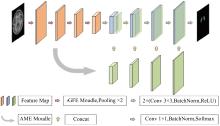
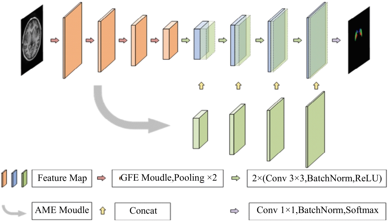
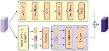
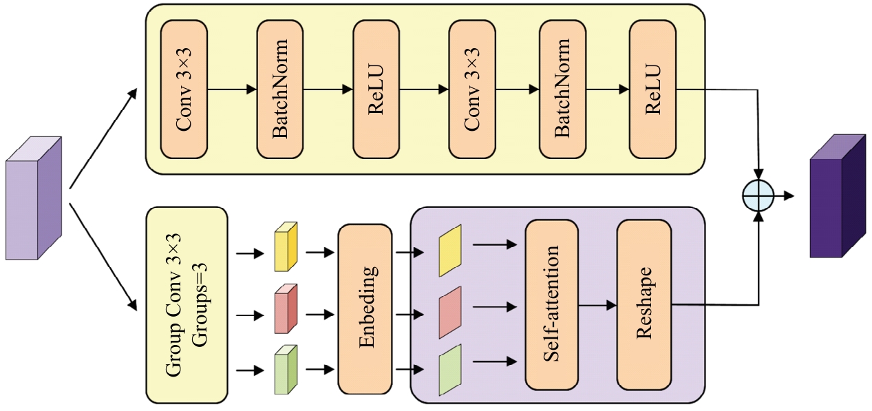
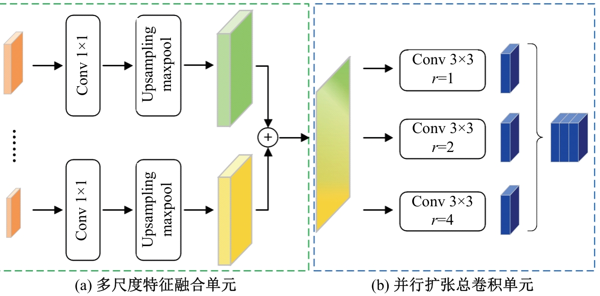
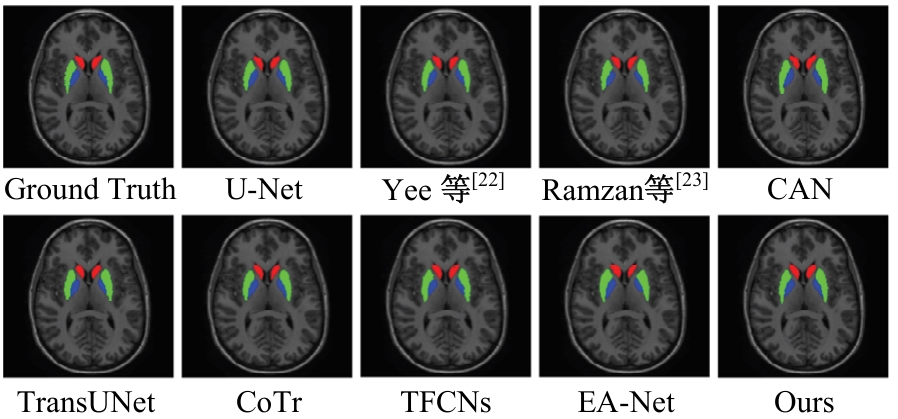
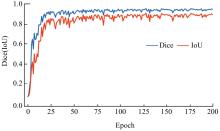
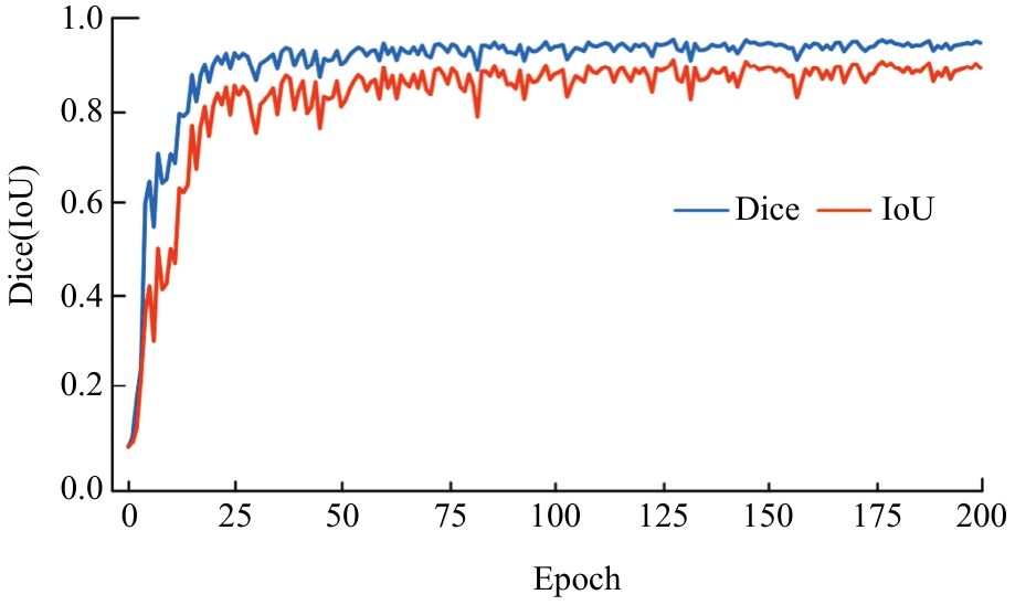
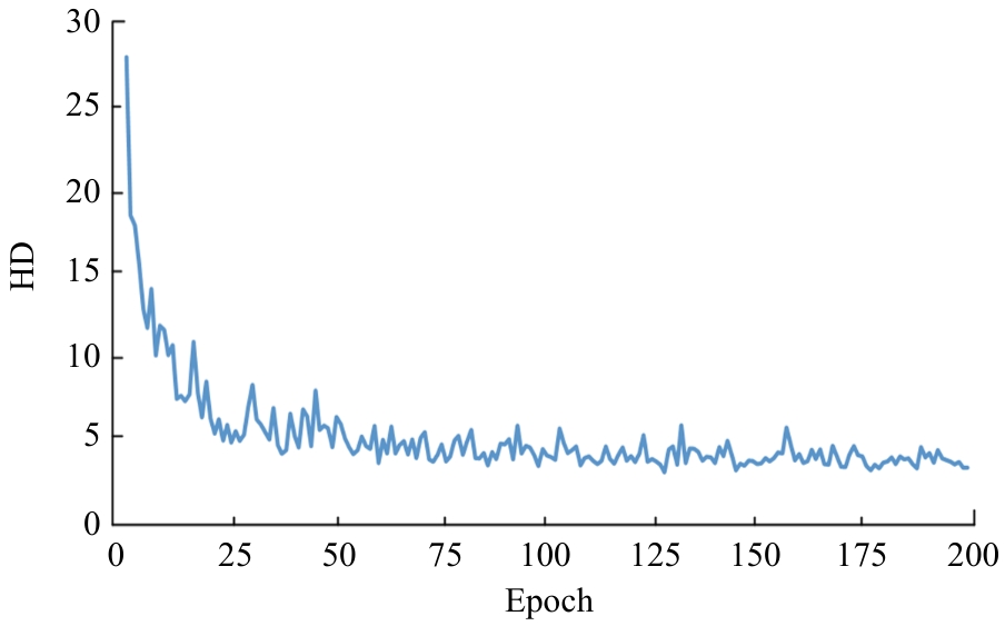
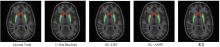
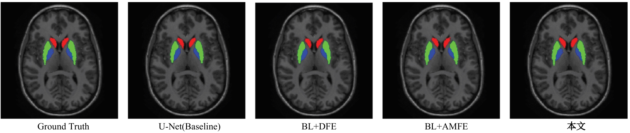
针对脑皮层下组织部分结构(如纹状体)在医学影像中目标小、对比度低,图像分割难度大,在自动医学诊断中应用比较困难的问题,本文基于深度学习的方法提出了一种医学图像分割网络,在磁共振成像中分割组成纹状体的苍白球、尾状核、壳核3部分。本文提出的网络模型具有捕获全局和局部特征的能力,并建立了全局与局部信息的相关性,在深度不退化的同时有效融合不同尺度的深层语义特征和浅层细节特征,实现对纹状体的精确分割。模型在公开的脑部数据集上进行了验证,并与其他先进的方法进行对比,结果表明本文的戴斯相似系数、平均交并比、95%豪斯多夫距离分别为94.26%、90.94%、3.82,均优于其他几种方法,达到了先进水平,这表明本文模型可以提高对纹状体的分割精度,为相关疾病的研究提供依据。
中图分类号:
- TP391.4

| [1] | Del Rey N L G, García-Cabezas M Á. Cytology, architecture, development, and connections of the primate striatum: hints for human pathology[J]. Neurobiology of Disease, 2023, 176: No. 105945. |
| [2] | Cataldi S, Stanley A T, Miniaci M C, et al. Interpreting the role of the striatum during multiple phases of motor learning[J]. The FEBS Journal, 2022, 289(8): 2263-2281. |
| [3] | Mätlik K, Baffuto M, Kus L, et al. Cell-type-specific CAG repeat expansions and toxicity of mutant Huntingtin in human striatum and cerebellum[J]. Nature Genetics, 2024, 56: 383-394. |
| [4] | Afrasiabi M, Redinbaugh M J, Phillips J M, et al. Consciousness depends on integration between parietal cortex, striatum, and thalamus[J]. Cell Systems, 2021, 12(4): 363-373. |
| [5] | Blumenstock S, Dudanova I. Cortical and striatal circuits in Huntington´s disease[J]. Frontiers in Neuroscience, 2020, 14: No. 82. |
| [6] | Valjent E, Gangarossa G. The tail of the striatum: from anatomy to connectivity and function[J]. Trends in Neurosciences, 2021, 44(3): 203-214. |
| [7] | Liu X, Song L, Liu S, et al. A review of deep-learning-based medical image segmentation methods[J]. Sustainability, 2021, 13(3): No.1224. |
| [8] | 刘近贞, 高国辉, 熊慧. 用于脑组织分割的多尺度注意网络[J]. 吉林大学学报: 工学版, 2023, 53(2): 576-583. |
| Liu Jin-zhen, Gao Guo-hui, Xiong Hui. Multi⁃scale attention network for brain tissue segmentation[J]. Journal of Jilin University(Engineering and Technology Edition), 2023, 53(2): 576-583. | |
| [9] | Jha D, Riegler M A, Johansen D, et al. Doubleu-net: a deep convolutional neural network for medical image segmentation[C]∥IEEE 33rd International Symposium on Computer-Based Medical Systems (CBMS), Rochester, USA, 2020: 558-564. |
| [10] | Lecun Y, Bottou L, Bengio Y, et al. Gradient-based learning applied to document recognition[J]. Proceedings of the IEEE, 1998, 86(11): 2278-2324. |
| [11] | Long J, Shelhamer E, Darrell T. Fully convolutional networks for semantic segmentation[C]∥Proceedings of the IEEE Conference on Computer Vision and Pattern Recognition, 2015: 3431-3440. |
| [12] | Ronneberger O, Fischer P, Brox T. U-net: convolutional networks for biomedical image segmentation[C]∥ The 18th International Conference on Medical Image Computing and Computer-Assisted Intervention, Munich, Germany, 2015: 234-241. |
| [13] | Chen L C, Papandreou G, Kokkinos I, et al. Semantic image segmentation with deep convolutional nets and fully connected crfs[J/OL].[2023-11-22]. . |
| [14] | Chen L C, Papandreou G, Kokkinos I, et al. Deeplab: semantic image segmentation with deep convolutional nets, atrous convolution, and fully connected crfs[J].IEEE Transactions on Pattern Analysis and Machine Intelligence, 2017, 40(4): 834-848. |
| [15] | Chen L C, Papandreou G, Schroff F, et al. Rethinking atrous convolution for semantic image segmentation[J/OL].[2023-11-23]. . |
| [16] | Chen L C, Zhu Y, Papandreou G, et al. Encoder-decoder with atrous separable convolution for semantic image segmentation[C]∥Proceedings of the European Conference on Computer Vision(ECCV), Munich, Germany, 2018: 801-818. |
| [17] | Safavian N, Batouli S A H, Oghabian M A. An automatic level set method for hippocampus segmentation in MR images[J]. Computer Methods in Biomechanics and Biomedical Engineering: Imaging & Visualization, 2020, 8(4): 400-410. |
| [18] | 黄鸿, 吕容飞, 陶俊利, 等. 基于改进 U-Net++ 的 CT 影像肺结节分割算法[J]. 光子学报, 2021, 50(2): 65-75. |
| Huang Hong, Rong-fei Lü, Tao Jun-li, et al. Segmentation of lung nodules in CT images using improved UNet++[J]. Acta Photonica Sinica, 2021, 50(2):65-75. | |
| [19] | Qin D, Bu J J, Liu Z, et al. Efficient medical image segmentation based on knowledge distillation[J]. IEEE Transactions on Medical Imaging, 2021, 40(12): 3820-3831. |
| [20] | Dosovitskiy A, Beyer L, Kolesnikov A, et al. An image is worth 16x16 words: Transformers for image recognition at scale[J/OL].[2023-11-24]. . |
| [21] | Vaswani A, Shazeer N, Parmar N, et al. Attention is all you need[C]∥Proceedings of the 31st International Conference on Neural Information Processing Systems,Long Beach California USA,2017: 6000-6010. |
| [22] | Yee E, Ma D, Popuri K, et al. 3D hemisphere-based convolutional neural network for whole-brain MRI segmentation[J]. Computerized Medical Imaging and Graphics, 2022, 95: No.102000. |
| [23] | Ramzan F, Khan M U G, Iqbal S, et al. Volumetric segmentation of brain regions from MRI scans using 3D convolutional neural networks[J]. IEEE Access, 2020, 8: 103697-103709. |
| [24] | Wu W, Gao L, Duan H, et al. Segmentation of pulmonary nodules in CT images based on 3D‐UNET combined with three‐dimensional conditional random field optimization[J]. Medical Physics, 2020, 47(9): 4054-4063. |
| [25] | Yan H, Chen A. A novel improved brain tumor segmentation method using deep learning network[J].Journal of Physics: Conference Series, 2021, 1944(1): No.012011. |
| [26] | Li Z, Zhang C, Zhang Y, et al. CAN: context-assisted full attention network for brain tissue segmentation[J]. Medical Image Analysis, 2023, 85: No.102710. |
| [27] | Xie Y, Zhang J, Shen C, et al. Cotr: efficiently bridging cnn and transformer for 3d medical image segmentation[C]∥The 24th International Conference on Medical Image Computing and Computer Assisted Intervention, Strasbourg, France,2021: 171-180. |
| [28] | Chen J, Lu Y, Yu Q, et al. Transunet: Transformers make strong encoders for medical image segmentation[J/OL].[2023-10-25]. . |
| [29] | Li Z, Li D, Xu C, et al. TFCNs: a CNN-transformer hybrid network for medical image segmentation[C]∥International Conference on Artificial Neural Networks,Bristol, UK, 2022: 781-792. |
| [30] | Wang K, Zhang X, Zhang X, et al. EANet: iterative edge attention network for medical image segmentation[J]. Pattern Recognition, 2022, 127: No.108636. |
| [1] | 姚宗伟,陈辰,高振云,靳鸿鹏,荣浩,李学飞,黄虹溥,毕秋实. 基于合成图像数据集的挖掘机关键点识别[J]. 吉林大学学报(工学版), 2026, 56(1): 76-85. |
| [2] | 王琳虹,刘宇阳,刘子昱,鹿应佳,张宇恒,黄桂树. 基于YOLOv5的轻量化桥梁缺陷识别[J]. 吉林大学学报(工学版), 2025, 55(9): 2958-2968. |
| [3] | 廉敬,张继保,刘冀钊,张家骏,董子龙. 基于文本引导的人脸图像修复[J]. 吉林大学学报(工学版), 2025, 55(8): 2732-2740. |
| [4] | 刘元宁,王星喆,黄子彧,张家晨,刘震. 基于多模态数据融合的胃癌患者生存预测模型[J]. 吉林大学学报(工学版), 2025, 55(8): 2693-2702. |
| [5] | 袁靖舒,李武,赵兴雨,袁满. 基于BERTGAT-Contrastive的语义匹配模型[J]. 吉林大学学报(工学版), 2025, 55(7): 2383-2392. |
| [6] | 徐慧智,郝东升,徐小婷,蒋时森. 基于深度学习的高速公路小目标检测算法[J]. 吉林大学学报(工学版), 2025, 55(6): 2003-2014. |
| [7] | 张汝波,常世淇,张天一. 基于深度学习的图像信息隐藏方法综述[J]. 吉林大学学报(工学版), 2025, 55(5): 1497-1515. |
| [8] | 李健,刘欢,李艳秋,王海瑞,关路,廖昌义. 基于THGS算法优化ResNet-18模型的图像识别[J]. 吉林大学学报(工学版), 2025, 55(5): 1629-1637. |
| [9] | 文斌,丁弈夫,杨超,沈艳军,李辉. 基于自选择架构网络的交通标志分类算法[J]. 吉林大学学报(工学版), 2025, 55(5): 1705-1713. |
| [10] | 赵宏伟,周明珠,刘萍萍,周求湛. 基于置信学习和协同训练的医学图像分割方法[J]. 吉林大学学报(工学版), 2025, 55(5): 1675-1681. |
| [11] | 李振江,万利,周世睿,陶楚青,魏巍. 基于时空Transformer网络的隧道交通运行风险动态辨识方法[J]. 吉林大学学报(工学版), 2025, 55(4): 1336-1345. |
| [12] | 赵孟雪,车翔玖,徐欢,刘全乐. 基于先验知识优化的医学图像候选区域生成方法[J]. 吉林大学学报(工学版), 2025, 55(2): 722-730. |
| [13] | 金虎,申玉生,方勇,于丽,周佳媚. 基于深度学习SSD算法的公路隧道衬砌细小裂缝识别[J]. 吉林大学学报(工学版), 2025, 55(11): 3653-3659. |
| [14] | 蔡晓东,黄业洋,董丽芳. 基于增强正例与层间负例的语义相似性模型[J]. 吉林大学学报(工学版), 2025, 55(11): 3705-3714. |
| [15] | 姜来为,王策,杨宏宇. 基于深度学习的多目标跟踪研究进展综述[J]. 吉林大学学报(工学版), 2025, 55(11): 3429-3445. |
|
||