吉林大学学报(医学版) ›› 2022, Vol. 48 ›› Issue (6): 1593-1598.doi: 10.13481/j.1671-587X.20220627

• 临床医学 • 上一篇    下一篇

肾脏孤立性纤维性肿瘤并发肾盂积水1例报告及文献复习

惠鹏祥,杨潇,王旭,张明,范海涛,于汇康,王寅春,赵群,汤高文,李然伟()   

  1. 吉林大学第二医院泌尿外科,吉林 长春 130041
  • 收稿日期:2021-12-15 出版日期:2022-11-28 发布日期:2022-12-07
  • 通讯作者: 李然伟 E-mail:ranwei1968@sina.com
  • 作者简介:惠鹏祥(1995-),男,陕西省榆林市人, 在读硕士研究生,主要从事肾脏疾病和肾上腺疾病基础及临床方面的研究。
  • 基金资助:
    吉林省科技厅发展计划项目(20191102012YY);吉林省财政厅卫生人才项目(2020SCZT056)

Renal solitary fibrous tumor complicated with hydronephrosis: A case report and literature review

Pengxiang HUI,Xiao YANG,Xu WANG,Ming ZHANG,Haitao FAN,Huikang YU,Yinchun WANG,Qun ZHAO,Gaowen TANG,Ranwei LI()   

  1. Department of Urology,Second Hospital,Jilin University,Changchun 130041,China
  • Received:2021-12-15 Online:2022-11-28 Published:2022-12-07
  • Contact: Ranwei LI E-mail:ranwei1968@sina.com

摘要:

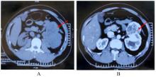
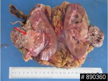
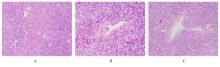
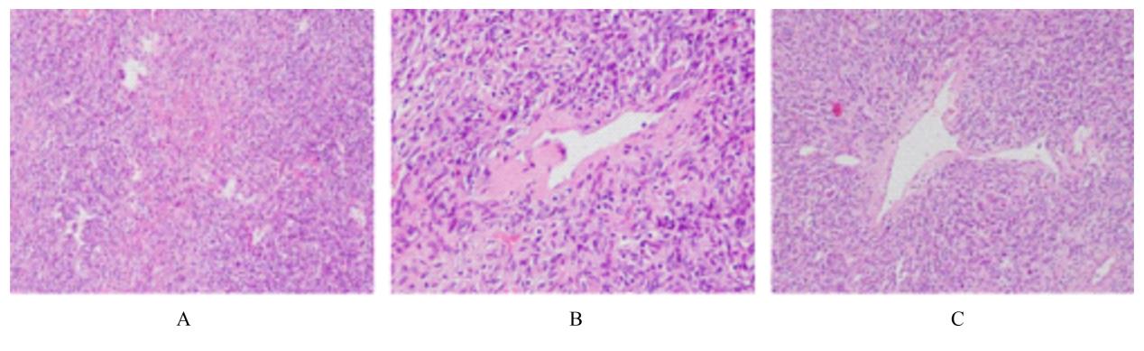
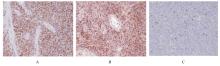
目的 分析肾脏SFT并发肾盂积水患者的临床表现、诊断和治疗方法,提高临床医生对该疾病的认识。 方法 收集1例肾脏SFT并发肾盂积水患者的临床症状、体征、影像学表现和术后病理结果等临床资料,并复习相关文献,总结肾脏SFT并发肾盂积水患者的临床特点、诊断和治疗方法。 结果 30岁男性患者因体检发现左侧肾肿物并发肾盂积水3 d入院。腹部超声提示左侧肾肿物和左侧肾盂积水,集合系统光点分离2.2 cm;进一步行CT检查显示左肾占位性病变并发肾盂积水,考虑肾透明细胞癌可能。初步诊断为左侧肾恶性肿瘤,术前准备后行经腹左侧根治性肾切除术;术后镜下见细胞丰富区和细胞稀疏区交替进行,梭形细胞呈无结构性排列。结合免疫组织化学染色结果[信号转导和转录激活因子6(STAT6)(+)、CD34(+)、Vimentin(+)、CD99(部分+)],确诊为肾脏SFT。术后患者恢复良好,切口甲级愈合,无并发症出院。术后1个月门诊复查全腹CT检查未见局部复发和转移的异常表现,术后3个月电话随访患者无不适症状。 结论 肾脏SFT无特异性临床表现,且容易误诊为其他肿瘤;确诊主要依赖形态学和免疫组织化学染色检查结果,目前手术治疗仍是肾脏SFT的首选治疗方法。

关键词: 孤立性纤维性肿瘤, 肾脏, 肾盂积水, 信号转导和转录激活因子6, 血管外皮瘤

Abstract:

Methods The clinical symptoms and signs, imaging findings and postoperative pathological results of one patient with renal SFT complicated with hydronephrosis were collected, the relevant literatures were reviewed,and the clinical characteristics, diagnosis and treatment of the patient with renal SFT complicated with hydronephrosis were summarized. Results A 30-year-old male patient was admitted to the hospital due to a left renal mass with hydronephrosis for 3 d detected by routine physical examination.The abdominal ultrasonography results revealed a left renal mass and left hydronephrosis, with a 2.2 cm separation of light spots in the collecting system; the CT examination results further revealed a left renal space-occupying lesion with hydronephrosis, and the possibility of renal clear cell carcinoma was considered. The preliminary diagnosis was the left renal malignancy, after preoperative preparation, the left transabdominal radical nephrectomy was performed;the postoperative microscope detection results showed that the cell-rich area and the cell-sparse area were alternated, and the spindle cells showed no structural arrangement. Combined with the immunohistochemical staining results [signal transducer and activator of transcription 6 (STAT6)(+),CD34(+),Vimentin(+),CD99 (partial+)],the diagnosis of renal SFT was confirmed. The patient recovered well after operation with grade A healing of the incision and was discharged without complications. The CT examination results showed that there was no abnormal manifestation of local recurrence and metastasis 1 month after operation, and no symptoms of discomfort were reported during the follow-up of 3 months after operation. Conclusion Renal SFT has no specific clinical manifestations and can be easily diagnosed as other tumors; the diagnosis mainly depends on the histopathology and immunohistochemical staining results,and currently surgery is still the first option for the treatment of renal SFT. Objective To analyze the clinical manifestations, diagnosis and treatment methods of the patient with renal solitary fibrous tumors (SFT) complicated with hydronephrosis,and to provide the understandings of the clinicians for SFT.

Key words: Solitary fibrous tumor, Kidney, Hydronephrosis, Signal transducer and activator of transcription 6, Hemangiopericytoma

中图分类号: 

  • R737.11