吉林大学学报(医学版) ›› 2023, Vol. 49 ›› Issue (6): 1593-1598.doi: 10.13481/j.1671-587X.20230624

• 临床医学 • 上一篇    下一篇

超声诊断十二指肠后壁穿孔致胆囊周围积液1例报告及文献复习

朱晴,吴明丽,刘琦尧,张镰竞,胡月,杨冬艳()   

  1. 吉林大学中日联谊医院超声科,吉林 长春 130033
  • 收稿日期:2023-01-16 出版日期:2023-11-28 发布日期:2023-12-22
  • 通讯作者: 杨冬艳 E-mail:yangdongy@jlu.edu.cn
  • 作者简介:朱 晴(1998-),女,山东省菏泽市人,在读硕士研究生,主要从事腹部超声诊断方面的研究。
  • 基金资助:
    吉林省科技厅科技发展计划项目(20200201554JC)

Ultrasonographic diagnosis of peri-gallbladder fluid caused by posterior duodenal perforation:A case report and literature review

Qing ZHU,Mingli WU,Qiyao LIU,Lianjing ZHANG,Yue HU,Dongyan YANG()   

  1. Department of Ultrasonography,China-Japan Union Hospital,Jilin University,Changchun 130033,China
  • Received:2023-01-16 Online:2023-11-28 Published:2023-12-22
  • Contact: Dongyan YANG E-mail:yangdongy@jlu.edu.cn

摘要:

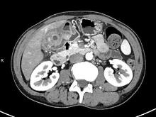
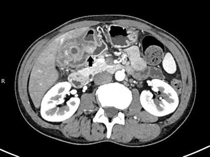
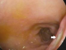
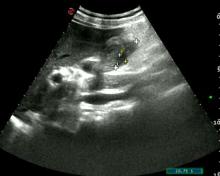
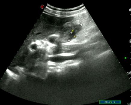
目的 分析1例由超声首诊的十二指肠后壁穿孔导致胆囊周围积液患者的影像学表现及其临床诊断和治疗经过,为该病的临床诊断提供依据。 方法 收集1例十二指肠后壁穿孔导致胆囊周围积液患者的临床资料、实验室检查、胃镜和影像学表现,记录其诊疗过程并进行随访。结合相关文献复习,分析十二指肠后壁穿孔的临床特点和影像学表现。 结果 患者,男性,50岁,无明显诱因右上腹持续性钝痛超过20 d、餐后加剧,同时伴有右腰背部放射性疼痛,于当地医院行超声检查提示胆囊占位性病变,为求进一步诊治就诊于本院。入院当日行腹部超声检查,可见空腹胆囊充盈极差,囊壁连续且弥漫均匀增厚,胆囊腔内见多发强回声,胆囊周围可见杂乱分布的无及弱回声延续至十二指肠球部后方,与球部以窄条状气体影相通。超声考虑为十二指肠球部后壁穿孔导致胆囊周围积液,胆囊受压充盈极差,胆囊壁慢性炎症改变伴胆囊结石。计算机断层扫描(CT)增强检查可见十二指肠球部后壁穿孔处清晰显示,CT结论与超声结论相似;胃镜检查显示十二指肠球部后壁深大溃疡,证实诊断为溃疡穿孔导致胆囊周围积液。置入胃管和空肠营养管,同时行消炎、镇痛和抑酸等对症治疗,治疗3个月内复查超声显示胆囊周围积液逐渐减少、胆囊腔逐渐充盈。患者因胆囊炎症状持续存在,遂行胆囊切除术,病理诊断为慢性胆囊炎并发胆囊结石。 结论 十二指肠后壁穿孔常因临床表现不典型而导致误诊或漏诊,影像学检查可辅助临床进行早期诊断,在腹部气体干扰未影响观察时,超声可作为消化道穿孔首选且有效的诊断方法。

关键词: 十二指肠后壁穿孔, 胆囊周围积液, 超声, 影像学检查, 消化道穿孔

Abstract:

Objective To analyze the imaging performance and clinical diagnosis and treatment process of one patient with peri-gallbladder fluid caused by the posterior duodenal perforation primarily diagnosed by ultrasound, and to provide the clinical diagnostic evidence for this disease. Methods The clinical data, laboratory examination, gastroscope performance, and imaging performance of one patient with peri-gallbladder fluid caused by the posterior duodenal perforation was collected. The process of diagnosis and treatment was recorded and followed up. The related literatures were reviewed to analyze the clinical characteristics and imaging performances of the posterior duodenal perforation. Results The patient was a 50-year-old male who had constant dull pain in the upper right abdomen for over 20 d, without obvious cause and intensified after meals, accompanied by radiculalgia in the right waist and back.The ultrasonic examination at a local hospital showed an occupying lesion in the gallbladder, and the patient came to our hospital for the further diagnosis and treatment. On the day of admission, the ultrasound examination showed poor fasting gallbladder filling, continuous and uniformly thickened gallbladder wall.The multiple strong echoes were observed in the gallbladder cavity, and the chaotic distributed hypoechoic and anechoic areas could be seen around the gallbladder, extending to the back of the duodenal bulb, and connected with the bulb by a narrow strip of gaseous shadow. The ultrasound results showed that there was perforation of the posterior wall of the duodenal bulb, leading to pericholecystic fluid accumulation, with poor gallbladder filling due to compression, and chronic inflammation of the gallbladder wall associated with gallstones. The enhanced computed tomography (CT) results clearly showed the perforation site in the posterior wall of the duodenal bulb, and the CT conclusion was consistent with the ultrasound conclusion. The gastroscope results showed a large deep ulcer on the posterior wall of the duodenal bulb, and it confirmed to be diagnosed as the peri-gallbladder fluid caused by ulcer perforation. A gastric tube and jejunal nutrition tube were inserted, and the symptomatic treatments such as anti-inflammation, analgesia, and acid suppression were carried out. The ultrasound reexamination within 3 months of treatment showed the peri-gallbladder fluid was gradually decreased and the gallbladder cavity was gradually filled.Since the patient’s gallbladder inflammation symptoms persisted,the cholecystectomy was performed, and it was pathologically diagnosed as chronic cholecystitis complicated with gallstones. Conclusion The posterior duodenal perforation is often misdiagnosed or missed due to atypical clinical manifestations.The imaging examination can assist in early clinical diagnosis. When the presence of abdominal gas does not interfere with observation, the ultrasound can serve as the first choice and effective method for the diagnosis of gastrointestinal perforation.

Key words: Posterior duodenal perforation, Peri-gallbladder fluid, Ultrasound, Radiographic examination, Gastrointestinal perforation

中图分类号: 

  • R575.63