吉林大学学报(医学版) ›› 2022, Vol. 48 ›› Issue (6): 1437-1447.doi: 10.13481/j.1671-587X.20220609

• 基础研究 • 上一篇    下一篇

参红补血颗粒对气滞血瘀型血管内皮功能障碍小鼠血管内皮的保护作用及其机制

刘俊秀1,周佳1,律广富2,王雨辰1,庄雪峰1,赵嘉睿1,黄晓巍1(),李瑞丽1()   

  1. 1.长春中医药大学药学院临床药学与中药药理教研室,吉林 长春 130117
    2.长春中医药大学 吉林省人参研究科学院中药药理组,吉林 长春 130117
  • 收稿日期:2022-03-29 出版日期:2022-11-28 发布日期:2022-12-07
  • 通讯作者: 黄晓巍,李瑞丽 E-mail:15948000740@163.com;377311060@qq.com
  • 作者简介:刘俊秀(1997-),女,江苏省连云港市人,在读硕士研究生,主要从事药效物质基础及其机制方面的研究。
  • 基金资助:
    吉林省卫健委科研项目(2021JC078);吉林省教育厅科研项目(JJKH20220872KJ);吉林省发改委创新能力建设项目(2021C011)

Protective effect of Shenhong Buxue Granule on vascular endothelium of mice with vascular endothelial dysfunction of Qi stagnation and blood stasis type and its mechanism

Junxiu LIU1,Jia ZHOU1,Guangfu LYU2,Yuchen WANG1,Xuefeng ZHUANG1,Jiarui ZHAO1,Xiaowei HUANG1(),Ruili LI1()   

  1. 1.Department of Clinical Pharmacy and Pharmacology of Chinese Medicine,School of Pharmacy,Changchun University of Chinese Medicine,Changchun 130117,China
    2.Department of Pharmacology of Traditional Chinese Medicine,Jilin Ginseng Academy,Changchun University of Chinese Medicine,Changchun 130117,China.
  • Received:2022-03-29 Online:2022-11-28 Published:2022-12-07
  • Contact: Xiaowei HUANG,Ruili LI E-mail:15948000740@163.com;377311060@qq.com

摘要:

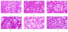
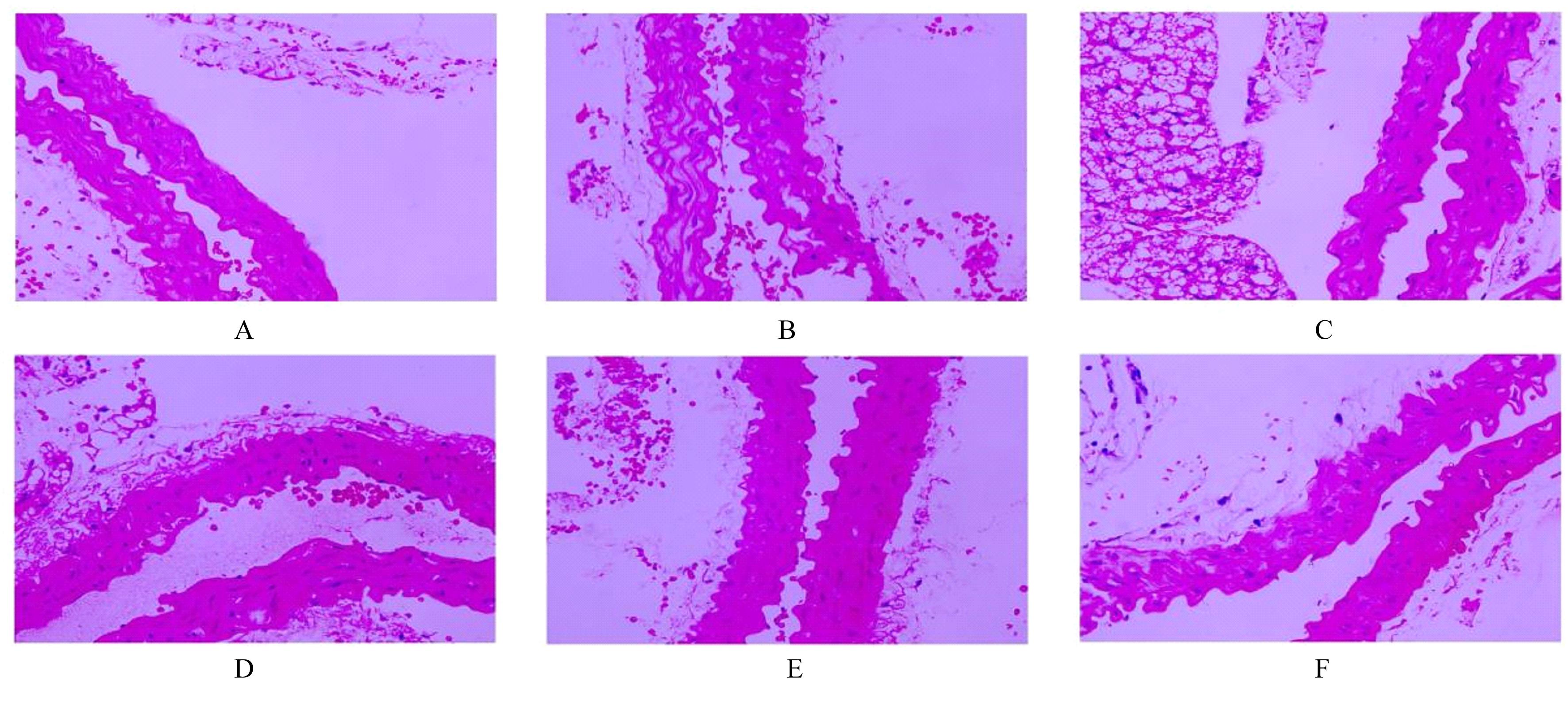
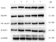
目的 探讨参红补血颗粒(SBG)对气滞血瘀型血管内皮功能障碍(VED)模型小鼠血管内皮的保护作用和对人脐静脉内皮细胞(HUVECs)中Toll样受体4(TLR4)、髓样分化因子(MyD88)、核因子 κBp65(NF-κB p65)及细胞间黏附分子1(ICAM-1)蛋白表达水平的影响,阐明其相关作用机制。 方法 将60只昆明小鼠随机分为正常对照组、VED组、阳性对照组(0.62 g·kg-1·d-1 血府逐瘀胶囊)、低剂量SBG(3 g·kg-1·d-1)组、中剂量SBG(6 g·kg-1·d-1)组和高剂量SBG(9 g·kg-1·d-1)组,每组10只。除正常对照组外,其他各组小鼠采用冰水浴游泳法构建气滞血瘀型VED模型,连续给药造模21 d。观察各组小鼠行为表现,血液流变仪检测各组小鼠全血黏度,苏木精-伊红(HE)染色观察各组小鼠肺和胸主动脉组织病理形态表现,ELISA法检测各组小鼠血清血管性血友病因子(vWF)、血栓调节蛋白(TM)、ICAM-1、肿瘤坏死因子α(TNF-α)和白细胞介素6(IL-6)水平,试剂盒检测各组小鼠血清一氧化氮(NO)水平和胸主动脉组织中谷胱甘肽过氧化物酶(GSH-Px)及超氧化物歧化酶(SOD)活性。体外培养人脐静脉内皮细胞(HUVECs),分为正常对照组,叔丁基过氧化氢(TBHP)组,低、中和高剂量SBG组,除正常对照组外,其他4组HUVECs采用TBHP诱导HUVECs建立氧化损伤模型,采用MTT法检测各组HUVECs存活率,Western blotting法检测各组HUVECs中TLR4、MyD88、NF-κB p65和ICAM-1蛋白表达水平。 结果 与正常对照组比较,VED组小鼠抓力值和自主活动次数明显降低(P<0.05), 全血黏度明显升高(P<0.05); 与VED组比较,阳性对照组和低、中及高剂量SBG组小鼠抓力值均明显升高(P<0.05),小鼠全血黏度明显降低(P<0.05),阳性对照组和高剂量SBG组小鼠自主活动次数明显升高(P<0.05)。HE染色,与正常对照组比较,VED组小鼠肺组织出现明显的毛细血管扩张和淤血,并且组织伴有大量炎性细胞浸润;与VED组比较,阳性对照组和不同剂量SBG组小鼠肺泡壁毛细血管扩张和淤血的程度减轻,炎性细胞浸润数量明显减少,并呈剂量依赖性;与正常对照组比较,VED组小鼠胸主动脉内壁结构紊乱,出现缺损和脱落;与VED组比较,阳性对照组和不同剂量SBG组小鼠血管内膜的脱落损伤情况改善,内膜相对完整。与正常对照组比较,VED组小鼠血清vWF、TM、ICAM-1、TNF-α和IL-6水平明显升高(P<0.05),血清NO水平以及动脉组织中GSH-Px和SOD活性明显降低(P<0.05);与VED组比较,阳性对照组和高剂量SBG组小鼠血清vWF、TM、ICAM-1、TNF-α和IL-6水平明显降低(P<0.05),血清NO水平以及动脉组织中GSH-Px和SOD活性明显升高(P<0.05)。与正常对照组比较,TBHP组HUVECs存活率明显降低(P<0.05),HUVECs中TLR4、MyD88、NF-κB p65和ICAM-1蛋白表达水平升高(P<0.05);与TBHP组比较,高剂量SBG组HUVECs存活率明显升高(P<0.05),HUVECs中TLR4、MyD88、NF-κB p65和ICAM-1蛋白表达水平明显降低(P<0.05)。 结论 SBG可通过抑制氧化应激,缓解炎症反应,达到保护气滞血瘀型VED小鼠血管内皮的作用。

关键词: 参红补血颗粒, 血管内皮功能障碍, 氧化应激, Toll样受体4, 人脐静脉内皮细胞

Abstract:

Objective To investigate the protective effect of Shenhong Buxue Granules (SBG) on vascular endothelium of the mice with vascular endothelial dysfunction (VED) of Qi stagnation and blood stasis type and its effect on the expression levels of Toll-like receptor 4 (TLR4),myeloid differentiation factor (MyD88),nuclear factor κB p65 (NF-κB p65),intercellular adhesion molecule-1 (ICAM-1) proteins in the human umbilical vein endothelial cells (HUVECs),and to elucidate its relevant action mechanism. Methods A total of 60 Kunming mice were randomly divided into normal control group, VED group, positive control group (0.62 g·kg-1·d-1 Xuefuzhuyu Capsule), low dose of SBG (3 g·kg-1·d-1) group, medium dose of SBG (6 g·kg-1·d-1) group and high dose of SBG (9 g·kg-1·d-1) group,and there were 10 mice in each group. Except for normal control group, the mice in other groups were used to construct the VED models of Qi stagnation and blood stasis type with ice water bath method,and the mice were continuously administered for 21 d. The behavior of the mice in various groups were observed,the whole blood viscosities of the mice in various groups were measured by blood rheometer,HE staining was used to observe the pathomorphology of lung and thoracic aorta tissues of the mice in various groups,the serum levels of von Willebrand factor (vWF), thrombomodulin (TM),ICAM-1, and tumor necrosis factor-α (TNF-α),and interleukin-6 (IL-6) of the mice in various groups were detected by ELISA method, and the kits were used to measure the levels of nitric oxide (NO) in serum and the activities of glutathione peroxidase (GSH-Px) and superoxide dismutase (SOD) in thoracic aorta tissue of the mice in various groups.The human umbilical vein endothelial cells (HUVECs) were cultured in vitro and divided into normal control group, tert-butyl hydroperoxide (TBHP) group and low,middle and high doses of SBG groups.Except for normal control group, the HUVECs in the other 4 groups were induced by TBHP to establish the oxidative damage model of HUVECs. The survival rates of HUVECs in various groups were detected by MTT assay, Western blotting method was used to detect the expression levels of TLR4, MyD88, NF-κB p65, and ICAM-1 proteins in the HUVECs in various groups. Results Compared with normal control group, the grasping force value and the number of autonomous activities of the mice in VED group were significantly decreased (P<0.05),and the whole blood viscosity was increased significantly (P<0.05); compared with VED group, the grasping force values of the mice in positive control group and the low, middle and high doses of SBG groups were significantly increased (P<0.05), the whole blood viscosities were significantly decreased (P<0.05),and the numbers of autonomous activities of the mice in positive control group and high dose of SBG group were significantly increased (P<0.05).The HE staining results showed that compared with normal control group, the lung tissue of the mice in VED group showed obvious telangiectasia and congestion, and the tissue was accompanied by a large number of inflammatory cell infiltration; compared with VED group, the degrees of telangiectasia and congestion of the alveolar wall of the mice in positive control group and different doses of SBG groups were decreased, and the number of inflammatory cell infiltration was significantly decreased in a dose-dependent manner; compared with normal control group, the inner wall of thoracic aorta of the mice in VED group was disordered, with defects and falling off; compared with VED group, the falling off lesion of vascular intima of the mice in positive control group and different doses of SBG groups was improved and the integrity of the intima was maintained. Compared with normal control group, the serum levels of vWF,TM,ICAM-1, TNF-α ,and IL-6 of the mice in VED group were significantly increased (P<0.05), and the serum level of NO and serum activities of GSH-Px and SOD in aorta tissue were significantly decreased (P<0.05); compared with VED group, the serum levels of vWF, TM, ICAM-1, TNF-α and IL-6 of the mice in positive control group and the high dose of SBG group were significantly decreased (P<0.05),and the serum level of NO and activities of GSH-Px and SOD in aorta tissue were significantly increased (P<0.05). Compared with normal control group, the survival rate of HUVECs in TBHP group was significantly decreased (P<0.05),and the expression levels of TLR4, MyD88, NF-κB p65, and ICAM-1 proteins in the HUVECs in TBHP group were significantly increased (P<0.05); compared with TBHP group, the survival rate of the HUVECs in high dose of SBG group was significantly increased (P<0.05),and the expression levels of TLR4, MyD88, NF-κB p65, and ICAM-1 proteins in the HUVECs in high dose of SBG group were significantly decreased (P<0.05). Conclusion SBG can protect the vascular endothelium of the mice with VED of Qi stagnation and blood stasis type by inhibiting the oxidative stress and alleviating the inflammatory response.

Key words: Shenhong Buxue Granules, Vascular endothelial dysfunction, Oxidative stress, Toll-like receptor 4, Human umbilical vein endothelial cells

中图分类号: 

  • R96