吉林大学学报(医学版) ›› 2022, Vol. 48 ›› Issue (6): 1448-1454.doi: 10.13481/j.1671-587X.20220610

• 基础研究 • 上一篇    下一篇

富血小板纤维蛋白和羟基磷灰石复合物的制备方法及其性能评价

张庆宇1,徐庭瑞1,矫君君2,夏德庚2,张天翼2,张莉1(),马宁2()   

  1. 1.吉林大学口腔医院急诊科,吉林 长春 130021
    2.吉林大学口腔医院牙周科,吉林 长春 130021
  • 收稿日期:2022-06-29 出版日期:2022-11-28 发布日期:2022-12-07
  • 通讯作者: 张莉,马宁 E-mail:zhang_li99@jlu.edu.cn;man@jlu.edu.cn
  • 作者简介:张庆宇(1997-),男,山东省淄博市人,在读硕士研究生,主要从事口腔软硬组织再生方面的研究。
  • 基金资助:
    吉林省教育厅科学技术研究规划项目(JJKH20211213KJ)

Preparation method of platelet-rich fibrin and hydroxyapatite complex and property evaluation

Qingyu ZHANG1,Tingrui XU1,Junjun JIAO2,Degeng XIA2,Tianyi ZHANG2,Li ZHANG1(),Ning MA2()   

  1. 1.Department of Emergency, Stomatology Hospital, Jilin University, Changchun 130021, China
    2.Department of Periodontics, Stomatology Hospital, Jilin University, Changchun 130021, China
  • Received:2022-06-29 Online:2022-11-28 Published:2022-12-07
  • Contact: Li ZHANG,Ning MA E-mail:zhang_li99@jlu.edu.cn;man@jlu.edu.cn

摘要:

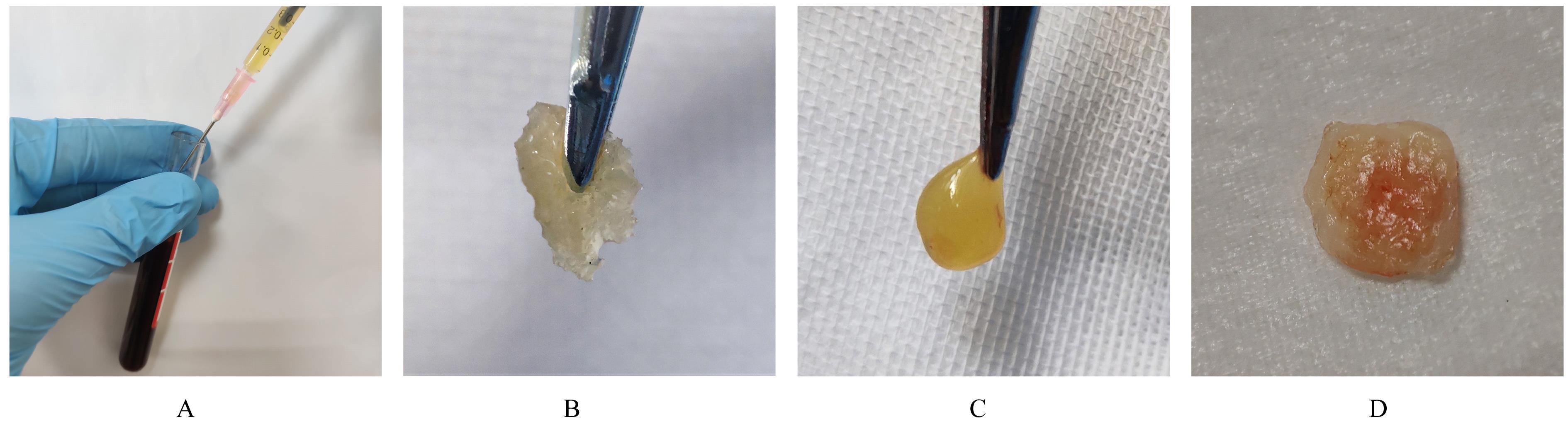
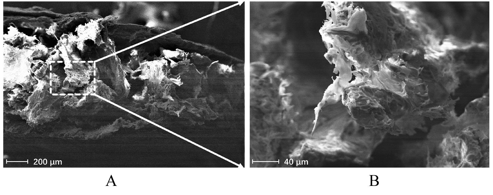
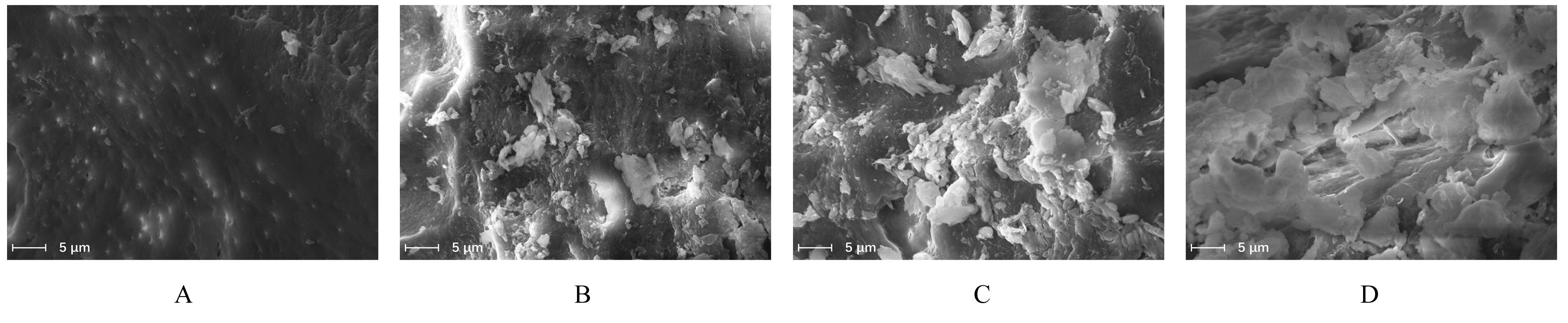
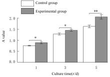
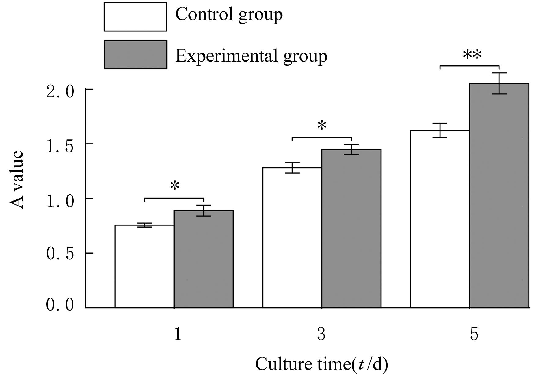
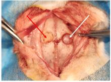
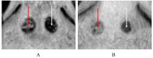
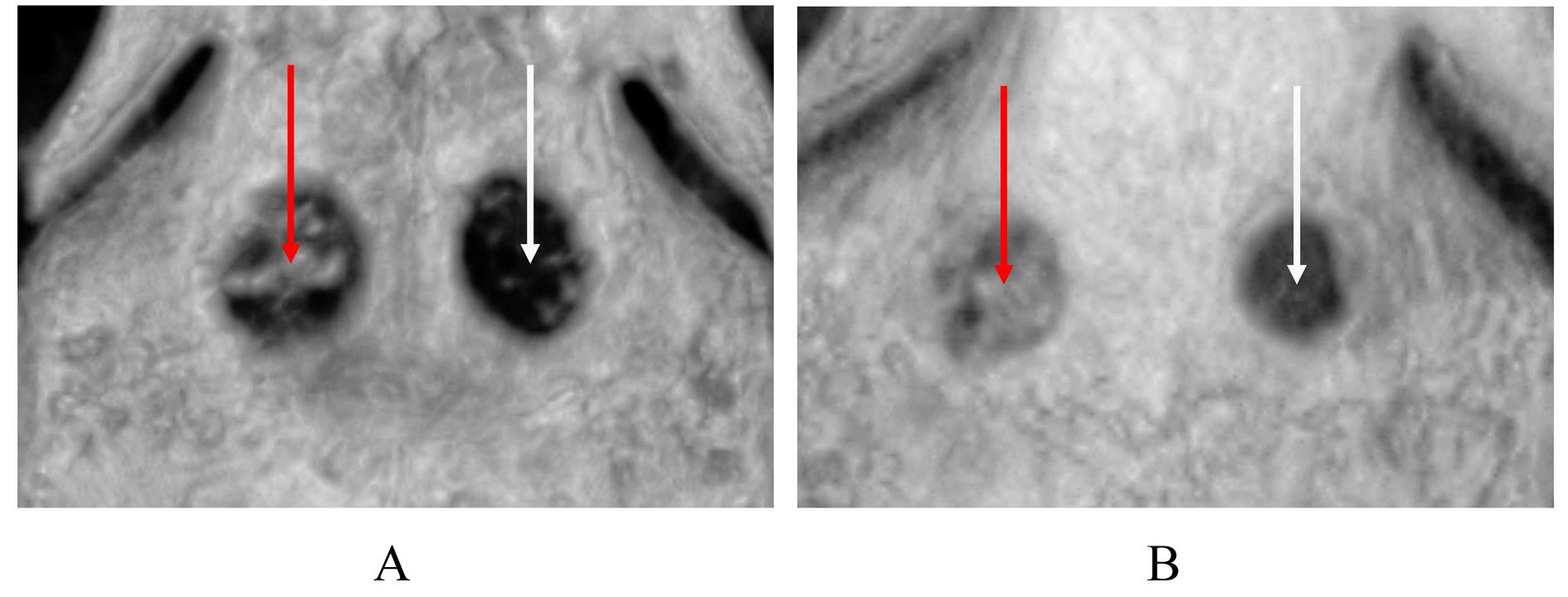
目的 探讨“三明治”法制备富血小板纤维蛋白(PRF)/羟基磷灰石(HA)复合物的工艺流程,并对其性能进行评价。 方法 采用700 r·min-1×3 min和1 300 r·min-1×14 min的离心参数,分别制备可注射型PRF(I-PRF)和改良型PRF(A-PRF),并采用“三明治”法将其与HA复合。冷冻干燥PRF/HA复合物后,扫描电子显微镜(SEM)观察其微观结构;模拟体液(SBF)浸泡材料,在第2、4、8和16天分别观察HA表面类骨层的生长情况,评价其矿化能力;采用PRF/HA复合物浸提液和完全培养基与小鼠前成骨MC3T3-E1细胞共培养作为实验组和对照组,在共培养第1、3和5天,CCK-8法检测各组前成骨MC3T3-E1细胞增殖活性。在日本大耳白兔的颅骨矢状缝两侧对称建立2个直径为6 mm的全层骨缺损区域,一侧不植入任何材料作为对照组,另一侧植入PRF/HA复合物作为实验组;术后4和8周时X射线平片及锥形束CT(CBCT)扫描术区,根据成像结果评价2组白兔体内的成骨效果。 结果 成功制备出具有“三明治”结构的PRF/HA复合物,外侧为A-PRF,中间为包裹了HA颗粒的I-PRF。SEM观察,复合物横断面为明显的三层结构,外侧为致密纤维蛋白网,内部为有纤维蛋白丝附着的多孔HA颗粒。PRF/HA复合物在SBF中浸泡后,第2天时HA表面即形成矿化结节,随着浸泡时间的延长,类骨层逐渐建立,第16天时已形成片层状结构。细胞增殖实验,与对照组比较,第1、3和5天时实验组MC3T3-E1细胞的增殖活性明显升高(P<0.05)。影像学观察,实验组颅骨缺损4周时已有明显的边缘成骨,8周时新生骨已几乎完全充填骨缺损区域,而对照组颅骨缺损新骨形成范围较实验组明显减少。 结论 “三明治”法制备的PRF/HA复合物,具有良好的矿化能力和生物相容性,且能够促进骨组织再生。

关键词: 富血小板纤维蛋白, 羟基磷灰石, 细胞增殖, 骨修复, 骨再生

Abstract:

Objective To discuss the technological process of platelet rich fibrin (PRF)/hydroxyapatite (HA) complex prepared by the “sandwich” method, and to evaluate its properties. Methods The injectable PRF (I-PRF) and advanced PRF (A-PRF) were prepared with the centrifugation indexes of 700 r·min-1×3 min and 1 300 r·min-1×14 min,respectively, then they were combined with HA using the “sandwich” method. The microstructure of PRF/HA complex was observed under scanning electron microscope (SEM) after freeze drying; the material was immersed in the simulated body fluid (SBF), and the growth of a bone-like layer on the surface of HA was observed on the 2nd, 4th, 8th,and 16th days, respectively,and its mineralization ability was evaluated;the PRF/HA complex extract solution co-cultured with preosteoblast MC3T3-E1 cells of the mice and complete culture medium were used as experimental group and control group;after cultured for 1, 3 and 5 d, respectively, the proliferation activities of the preosteoblast MC3T3-E1 cells were evaluated by CCK-8 method. Two full-thickness cranial defect areas with a diameter of 6 mm were symmetrically established on both the sides of the sagittal suture in the Japanese white rabbits; one side implanted with PRF/HA complex was regarded as experimental group, the other side without any material implantion was regarded as control group;X-ray and cone beam CT (CBCT) were used on the surgical area to evaluate the bone formation performance of the rabbits in two groups 4 and 8 weeks after surgery. Results The PRF/HA complex with a “sandwich” structure was successfully prepared, with the A-PRF on the outside and the I-PRF mixed with HA particles on the inside. The SEM observation results showed that the cross-section of the complex was an obvious three-layer structure, with the dense fibrin net on the outside and the porous HA particles attached by fibrin filaments on the inside. After being immersed in SBF,the mineralized nodules were formed on the surface of HA on the 2nd day, the bone-like layer was gradually established with the prolongation of the immersion time, and a lamellar structure was formed at the 16th day. The cell proliferation experiment results showed that compared with control group, the proliferation activities of the MC3T3-E1 cells in experimental group were significantly increased on the 1st, 3rd,and 5th days (P<0.05). The imaging observation results showed that the cranial defect in experimental group had obvious marginal osteogenesis 4 weeks after surgery and the new bone had almost completely filled the bone defect area 8 weeks after surgery, while the new bone formation of the cranial defect in control group was significantly decreased than that in experimental group. Conclusion PRF/HA complex prepared by the “sandwich” method has a good mineralization ability and biocompatibility, and can promote the bone regeneration.

Key words: Platelet rich fibrin, Hydroxyapatite, Cell proliferation, Bone repair, Bone regeneration

中图分类号: 

  • R782.1