吉林大学学报(医学版) ›› 2025, Vol. 51 ›› Issue (1): 9-16.doi: 10.13481/j.1671-587X.20250102

• 基础研究 • 上一篇    下一篇

低剂量甲氨蝶呤联合索拉非尼对小鼠骨肉瘤移植瘤的影响及其机制

王凤娇,顾超,胡沙,冯琴,郑儒娟,朱增燕(),王文娟()   

  1. 苏州大学附属儿童医院药剂科,江苏 苏州 215000
  • 收稿日期:2024-02-26 接受日期:2024-03-27 出版日期:2025-01-28 发布日期:2025-03-06
  • 通讯作者: 朱增燕,王文娟 E-mail:zhuzengyan7676@suda.edu.cn;wangwenjuan1110@163.com
  • 作者简介:王凤娇(1988-),女,江苏省南通市人,副主任药师,药理硕士,主要从事骨肉瘤治疗药物药理学方面的研究。
  • 基金资助:
    国家自然科学基金青年基金项目(32200775);江苏省教育厅高等学校基础科学(自然科学)面上项目(21KJB320017);苏州市科技局科教强卫项目(KJXW2020026)

Effect of low dose of methotrexate combined with sorafenib on osteosarcoma xenografts of mice and its mechanism

Fengjiao WANG,Chao GU,Sha HU,Qin FENG,Rujuan ZHENG,Zengyan ZHU(),Wenjuan WANG()   

  1. Department of Pharmacy,Affiliated Children’s Hospital,Soochow University,Suzhou 215000,China
  • Received:2024-02-26 Accepted:2024-03-27 Online:2025-01-28 Published:2025-03-06
  • Contact: Zengyan ZHU,Wenjuan WANG E-mail:zhuzengyan7676@suda.edu.cn;wangwenjuan1110@163.com

摘要:

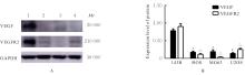
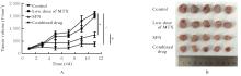
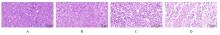
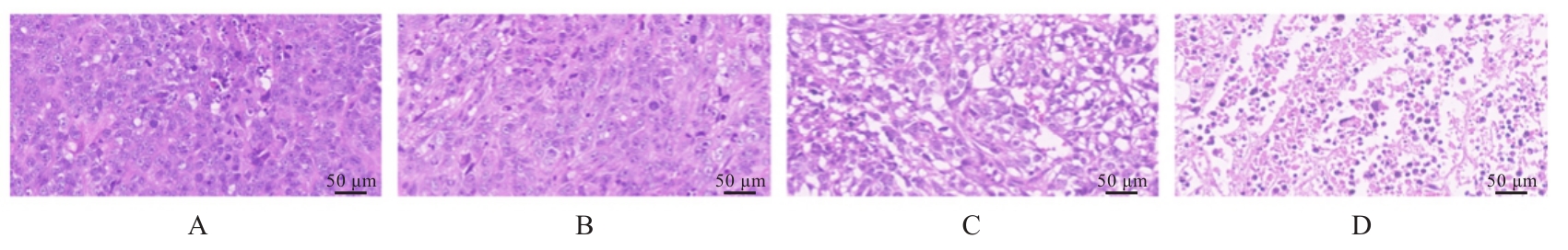
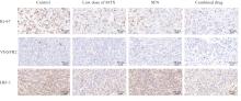
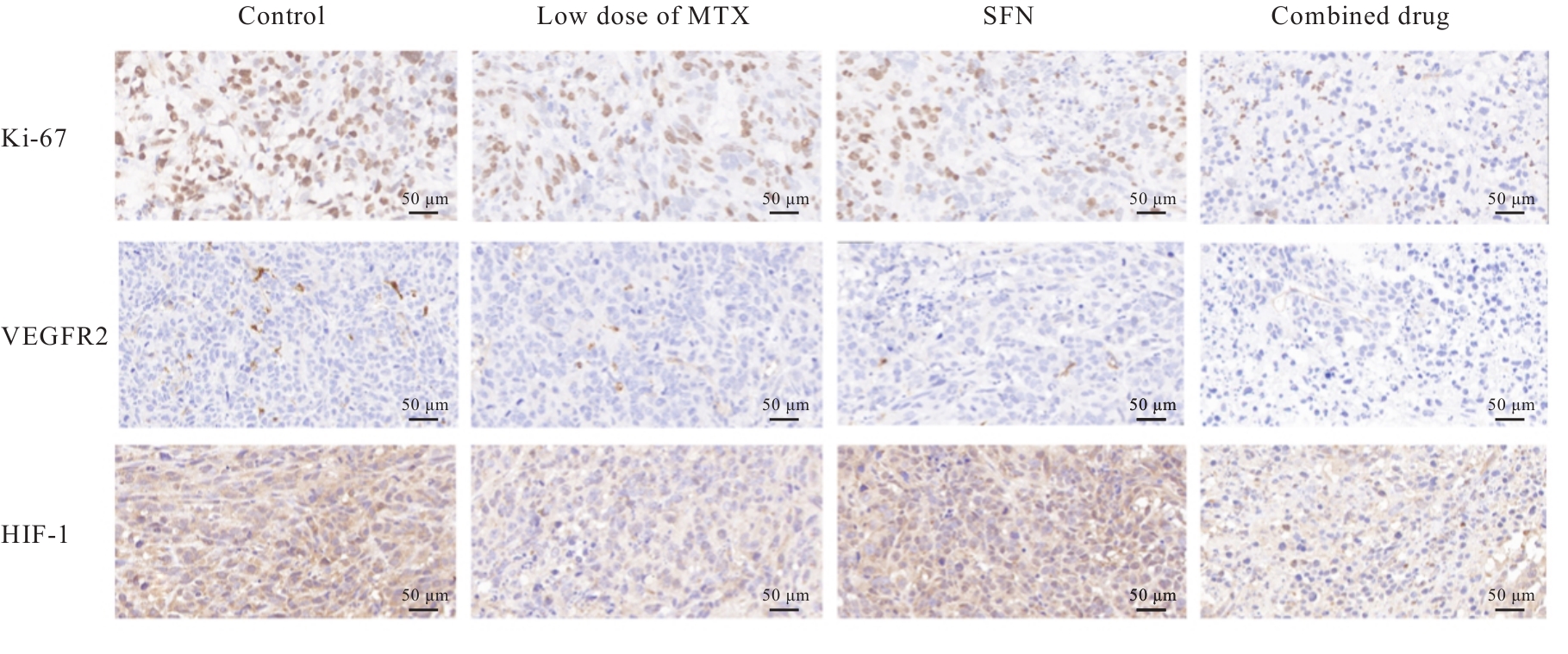
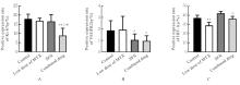
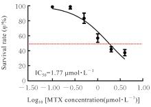
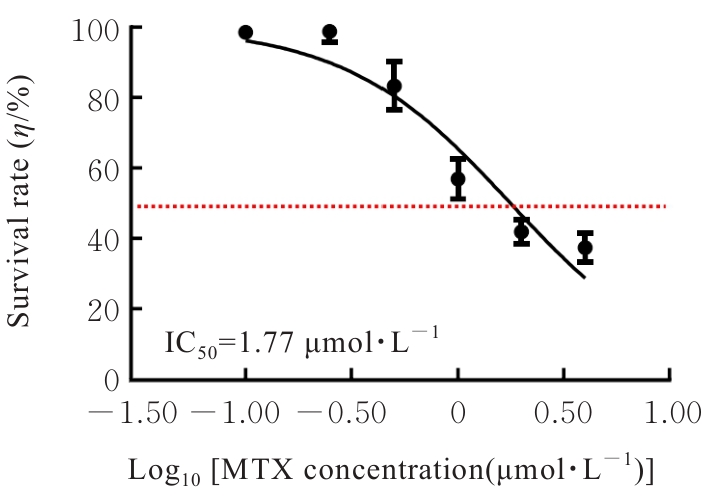
目的 探讨低剂量甲氨蝶呤(MTX)联合索拉非尼(SFN)对人骨肉瘤(OS)的抗肿瘤作用,并阐明其可能的作用机制。 方法 体外培养4种人OS细胞(143B细胞、HOS细胞、U2OS细胞和MG63细胞),采用Western blotting法测定各种细胞中的血管内皮生长因子(VEGF)和血管内皮细胞生长因子受体2(VEGFR2)蛋白表达水平;建立人OS裸鼠皮下移植瘤模型,并将建模成功的20只BABL/C裸鼠随机分为对照组(给予2%二甲基亚砜+98%玉米油)、低剂量MTX组(给予2 mg·kg-1 MTX)、SFN组(给予15 mg·kg-1 SFN)和联合用药组(给予2 mg·kg-1 MTX+ 15 mg·kg-1 SFN),每组5只,测量各组小鼠肿瘤体积并绘制肿瘤生长曲线;HE染色观察4组小鼠肿瘤组织病理形态表现,免疫组织化学法检测各组小鼠肿瘤组织中VEGFR2、细胞增殖抗原Ki-67和缺氧诱导因子1(HIF-1)蛋白阳性表达率;人OS 143B细胞分别给予0、0.125、0.250、0.500、1.000、2.000和4.000 μmol·L-1 MTX处理,CCK-8法检测各组143B细胞存活率,计算半数抑制浓度(IC50),并选取对143B细胞存活无影响的MTX浓度作为低剂量MTX;人OS 143B细胞分为对照组和低剂量MTX组(给予0和0.250 μmol·L-1 MTX处理),酶联免疫吸附试验(ELISA)法检测各组143B细胞中VEGF水平。 结果 与143B细胞比较,HOS细胞、U2OS细胞和MG63细胞中VEGF及VEGFR2蛋白表达水平明显降低(P<0.001)。裸鼠皮下移植瘤模型中,与对照组比较,SFN组和联合用药组小鼠皮下移植瘤体积减少(P<0.001),与低剂量MTX组和SFN组比较,联合用药组小鼠皮下移植瘤体积减少(P<0.01)。免疫组织化学法,与对照组比较,联合用药组小鼠肿瘤组织中Ki-67、VEGFR2和HIF-1蛋白阳性表达率明显降低(P<0.05)。CCK-8法,0.250 μmol·L-1 MTX对143B细胞增殖无明显改变。ELISA法,与对照组比较,低剂量MTX组143B细胞中VEGF水平明显降低(P<0.05)。 结论 低剂量MTX促进了SFN对人OS的抗肿瘤作用,其可能是通过抑制OS细胞分泌VEGF进而增强SFN对人OS的抗肿瘤作用。

关键词: 骨肉瘤, 索拉非尼, 甲氨蝶呤, 抗肿瘤, 血管内皮生长因子

Abstract:

Objective To discuss the anti-tumor effect of low dose of methotrexate(MTX) combined with sorafenib(SFN) on the human osteosarcoma(OS), and to clarify the possible mechanism. Methods Four types of human OS cells(143B cells, HOS cells, U2OS cells, and MG63 cells) were cultured in vitro. Western blotting method was used to detect the expression levels of vascular endothelial growth factor(VEGF) and vascular endothelial growth factor receptor 2(VEGFR2) proteins in the above four kinds of cells. The human OS xenograft model was established in the nude mice, and 20 successfully modeled BALB/C nude mice were randomly divided into control group(given 2% dimethyl sulfoxide+98% corn oil), low dose of MTX group(given 2 mg·kg-1 MTX), SFN group (given 15 mg·kg-1 SFN), and combined drug group(given 2 mg·kg-1 MTX+15 mg·kg-1 SFN); there were 5 mice in each group. The tumor volumes of the mice in various groups were detected and tumor growth curves were plotted; HE staining was used to observe the morphology of tumor tissue of the mice in various groups; immunohistochemistry was used to detect the positive expression rates of VEGFR2, proliferation marker Ki-67, and hypoxia-inducible factor-1(HIF-1) proteins in tumor tissue of the mice in various groups.The human OS 143B cells were divided into 0, 0.125, 0.250, 0.500, 1.000, 2.000, and 4.000 μmol·L-1 MTX groups(given 0, 0.125, 0.250, 0.500, 1.000, 2.000, and 4.000 μmol·L-1 MTX, respectively). CCK-8 method was used to detect the proliferation rates of the 143B cells in various groups, and half inhibityory concentration (IC50) was calculated; the concentration of MTX that had no effect on 143B cell survival was selected as low dose of MTX. The human OS 143B cells were divided into control and low dose of MTX groups (given 0 and 0.250 μmol·L-1 MTX). ELISA method was used to detect the levels of VEGF in the 143B cells in various groups. Results Compared with 143B cells, the expression levels of VEGF and VEGFR2 proteins in the HOS cells, U2OS cells, and MG63 cells were significantly increased (P<0.001). In the xenograft model, compared with control group, the tumor volumes of the mice in SFN group, and combined drug group were decreased (P<0.001); compared with low dose of MTX group and SFN group, the tumor volume of the mice in combined drug group was decreased (P<0.01). The immunohistochemical results showed that compared with control group, the positive expression rates of Ki-67, VEGFR2, and HIF-1 proteins in tumor tissue of the mice in combined drug group were significantly decreased(P<0.05). The CCK-8 results showed that there was no change in the proliferation of the 143B cells treated with 0.25 μmol·L-1 MTX. The ELISA results showed that compared with control group, the level of VEGF in the 143B cells in MTX group was signyicantly decreased (P<0.05). Conclusion Low dose of MTX enhances the anti-tumor effect of SFN on the human OS, which may be due to the inhibition of VEGF secretion by the OS cells, thereby enhancing the anti-tumor effect of SFN on the human OS.

Key words: Osteosarcoma, Sorafenib, Methotrexate, Anti-tumor, Vascular endothelial growth factor

中图分类号: 

  • R738.1