吉林大学学报(医学版) ›› 2025, Vol. 51 ›› Issue (4): 887-895.doi: 10.13481/j.1671-587X.20250404

• 基础研究 • 上一篇    下一篇

草苁蓉环烯醚萜苷对大鼠肝脏癌前病变的抑制作用及其机制

许惠仙1,徐慧2,全吉淑2(),郑峰1,3()   

  1. 1.延边大学附属医院临床营养科,吉林 延吉 133000
    2.延边大学医学院生物化学与分子生物学 教研室,吉林 延吉 133002
    3.山东省临沂市人民医院肿瘤科,山东 临沂 276000
  • 收稿日期:2024-08-23 接受日期:2024-10-20 出版日期:2025-07-28 发布日期:2025-08-25
  • 通讯作者: 全吉淑,郑峰 E-mail:quanjs@ybu.edu.cn;zhengfeng008@126.com
  • 作者简介:许惠仙(1982-),女,吉林省延吉市人,副主任医师,医学博士,主要从事分子药理学方面的研究。
  • 基金资助:
    国家自然科学基金项目(82060113);国家自然科学基金项目(81760659)

Inhibitory effect of iridoid glycosides from Boschniakia rossica on hepatic preneolasia of rats and its mechanism

Huixian XU1,Hui XU2,Jishu QUAN2(),Feng ZHENG1,3()   

  1. 1.Department of Clinical Nutrition,Affilited Hospital,Yanbian University,Yanji 133002,China
    2.Department of Biochemistry and Molecular Biology,School of Medical Sciences,Yanbian University,Yanji 133002,China
    3.Department of Oncology,People’s Hosptial,Linyi City,Shangdong Province,Linyi 276000,China
  • Received:2024-08-23 Accepted:2024-10-20 Online:2025-07-28 Published:2025-08-25
  • Contact: Jishu QUAN,Feng ZHENG E-mail:quanjs@ybu.edu.cn;zhengfeng008@126.com

摘要:

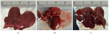
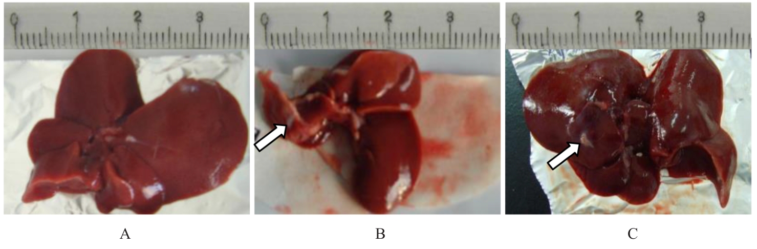
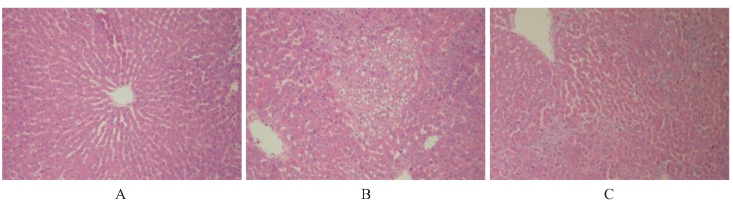
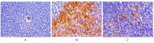
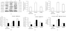
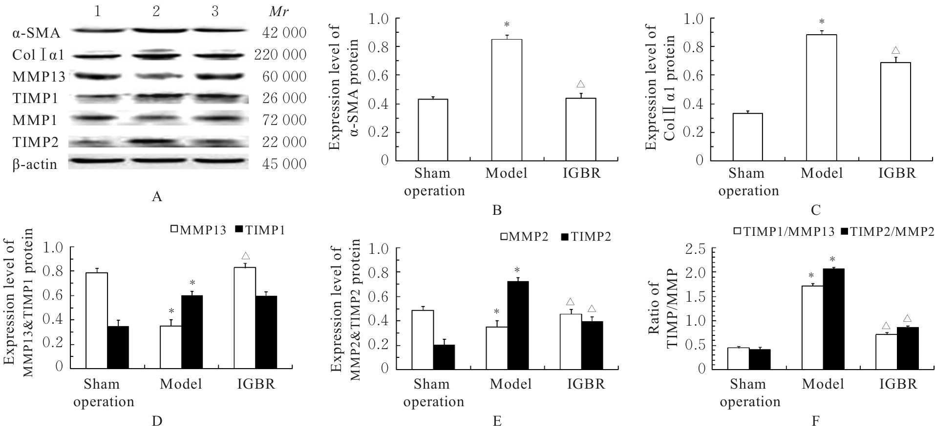
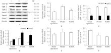
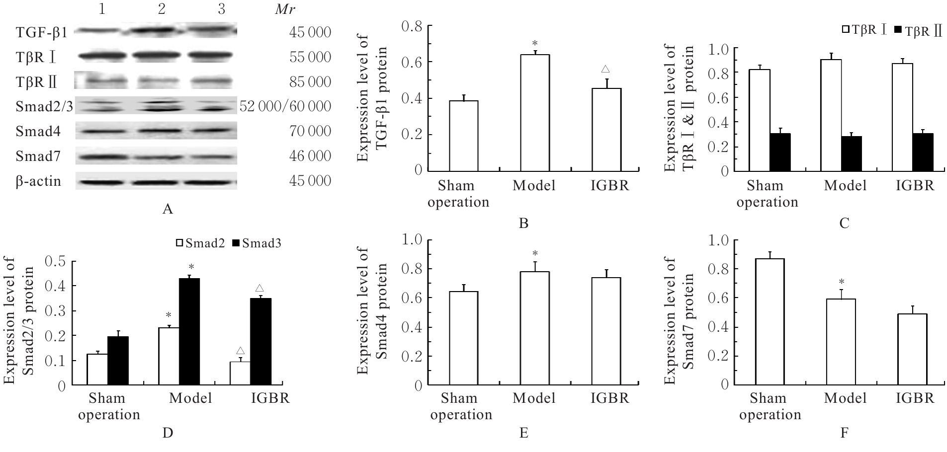
目的 研究草苁蓉环烯醚萜苷(IGBR)对大鼠肝脏癌前病变的防治作用,并阐明其可能的作用机制。 方法 选取30只Wistar大鼠,按照改良Solt-Faber法构建大鼠肝脏癌前病变模型。将制模成功后的大鼠随机分为假手术组、模型组和IGBR组,每组10只。取各组大鼠肝组织观察其形态表现,检测各组大鼠肝脏质量、肝脏指数和肝脏再生度,HE染色法观察各组大鼠肝组织病理形态表现,免疫组织化学法检测各组大鼠肝组织中谷胱甘肽-S-转移酶(GST)-Pi蛋白表达情况,比色法检测各组大鼠肝组织和肝线粒体中γ-谷氨酰转肽酶(γ-GT)、过氧化氢酶(CAT)、超氧化物岐化酶(SOD)、谷胱甘肽过氧化物酶(GSH-Px)和GST活性及丙二醛(MDA)水平,Western blotting法检测各组大鼠肝组织中α-平滑肌肌动蛋白(α-SMA)、Ⅰ型胶原蛋白α1链(ColⅠα1)、基质金属蛋白酶(MMP)13、MMP2、组织金属蛋白酶抑制剂(TIMP)1、TIMP2、转化生长因子β1(TGF-β1)、TGF-β1受体(TβR)、抗DPP同源物(Smad)2/3、Smad4和Smad7蛋白表达水平。 结果 与假手术组比较,模型组大鼠肝脏质量和肝脏指数升高(P<0.05);与模型组比较,IGBR组大鼠肝脏质量、肝脏指数和肝再生度均有下降趋势,但差异无统计学意义(P>0.05)。HE染色法,假手术组大鼠肝小叶结构完整清晰,肝细胞体积大、胞质丰富、嗜酸性,肝细胞以中央静脉为中心单行排列呈条索状向四周放射状排列,肝板间有不规则肝窦,仅于汇管区有少许胶原纤维存在和少量的炎症细胞浸润,肝细胞无变性坏死;模型组大鼠肝细胞失去正常排列,肝小叶结构消失,汇管区有以卵圆细胞为主的小细胞增生,纤维隔内有大量胶原沉积,有较明显纤维组织增生,肝细胞胞浆疏松,发生广泛的变性水肿,水样变性、气球样变性甚至灶状坏死,肝小叶中见增生的嗜碱性肝细胞形成细胞增生区,其细胞胞浆透亮,细胞核位于细胞中央,体积不大,染色质丰富,有1或2个明显核仁,肝小叶内还可见体积增大,细胞核和细胞浆着色浅而透明呈毛玻璃样的透明肝细胞灶;IGBR组大鼠肝小叶结构基本存在,可见肝细胞炎性病变减轻,水肿较轻,可见较多点状坏死或灶状坏死,核异型性大,可见病理性核分裂象或双核。免疫组织化学法,GST-Pi蛋白阳性灶为胞浆染色,呈棕黄色的圆形或类圆形结节;模型组大鼠肝组织中可观察到GST-Pi蛋白阳性灶,提示大鼠肝脏癌前病变模型制作成功。IGBR组大鼠肝组织中可观察到散在的GST-Pi蛋白阳性灶,较模型组明显减少。与假手术组比较,模型组大鼠肝组织中γ-GT活性升高(P<0.05);与模型组比较,IGBR组大鼠肝组织中γ-GT活性降低(P<0.05)。与假手术组比较,模型组大鼠肝组织和肝线粒体中GST活性及MDA水平升高(P<0.05),SOD、CAT和GSH-Px活性降低(P<0.05);与模型组比较,IGBR组大鼠肝组织和肝线粒体中GST活性及MDA水平降低(P<0.05),SOD、CAT和GSH-Px活性升高(P<0.05)。Western blotting法,与假手术组比较,模型组大鼠肝组织中α-SMA、ColⅠα1、TIMP1和TIMP2蛋白表达水平升高(P<0.05),MMP13和MMP2蛋白表达水平降低(P<0.05),TIMP1/MMP13和TIMP2/MMP2比值升高(P<0.05);与模型组比较,IGBR组大鼠肝组织中α-SMA、ColⅠα1和TIMP2蛋白表达水平降低(P<0.05),MMP13和MMP2蛋白表达水平升高(P<0.05),TIMP1/MMP13和TIMP2/MMP2比值降低(P<0.05)。与假手术组比较,模型组大鼠肝组织中TGF-β1、Smad2/3和Smad4蛋白表达水平升高(P<0.05);与模型组比较,IGBR组大鼠肝组织中TGF-β1、Smad2/3和Smad7蛋白表达水平降低(P<0.05)。 结论 IGBR对大鼠肝脏癌前病变和肝纤维化具有抑制作用,其机制可能与其增强肝组织抗氧化能力、抑制TGF-β/Smad信号通路及调控TIMP/MMP平衡有关。

关键词: 草苁蓉环烯醚萜苷, 肝脏癌前病变, 肝纤维化, 转化生长因子β1, 超氧化物岐化酶

Abstract:

Objective To investigate the preventive and therapeutic effects of iridoid glycosides from Boschniakia rossica (IGBR) on precancerous lesions of liver cancer in rats, and to clarify its possible mechanism. Methods Thirty Wistar rats were selected and the precancerous liver lesion model was established using the modified Solt-Faber method. The rats were randomly divided into sham operation group, model group, and IGBR group, and there were 10 rats in each group. The morphology of liver tissue of the rats in various groups were observed; the liver weights, liver indexes and liver regeneration degrees of the rats in various groups were measured; HE staining was used to observe the pathomorphology of liver tissue of the rats in various groups; immunohistochemistry method was used to detect the expression of glutathione-S-transferase(GST)-Pi protein in liver tissue of the rats in various groups; colorimetric method was used to detect the activities of γ-glutamyl transpeptidase (γ-GT), catalase (CAT), superoxide dismutase (SOD), glutathione peroxidase (GSH-Px) and glutathione S-transferase (GST), and the level of malondialdehyde (MDA) in liver tissue and mitochondria of the rats in various groups; Western blotting method was used to detect the expression levels of α-smooth muscle actin (α-SMA), collagen type Ⅰ alpha 1 chain (ColⅠα1), matrix metalloproteinase (MMP) 13, MMP2, tissue inhibitor of metalloproteinase (TIMP) 1, TIMP2, transforming growth factor β1 (TGF-β1), TGF-β1 receptor (TβR), mothers against decapentaplegic homolog (Smad) 2/3, Smad4, and Smad7 proteins in liver tissue of the rats in various groups. Results Compared with sham operation group, the liver weight, liver index and degree of liver regeneration of the rats in model group were increased (P<0.05); compared with model group, the liver weight and liver index of the rats in IGBR group showed a decreasing trend, but the difference was not statistically significant (P>0.05). The HE staining results showed that the liver lobule structure in sham operation group was intact and clear, with large hepatocytes, abundant eosinophilic cytoplasm, and hepatocytes arranged in single cords radiating from the central vein; there were irregular hepatic sinusoids between plates, with only a small amount of collagen fibers and inflammatory cell infiltration in the portal area, and no degeneration or necrosis of hepatocytes. In model group, the normal arrangement of hepatocytes disappeared, the liver lobule structure was disrupted, small cell hyperplasia (mainly oval cells) was observed in the portal area, with massive collagen deposition and significant fibrous tissue hyperplasia in the fibrous septum; hepatocytes showed extensive degenerative edema, hydropic degeneration or even ballooning degeneration and focal necrosis; basophilic hepatocytes formed proliferative areas with clear cytoplasm, centrally located nuclei and 1-2 prominent nucleoli; glassy hepatocytes with enlarged nuclei and pale transparent cytoplasm were also observed. In IGBR group, the liver lobule structure was basically preserved, with reduced inflammatory lesions, mild edema, and scattered spotty or focal necrosis; nuclear atypia and pathological mitotic figures or binucleation were observed. The immunohistochemistry results showed GST-Pi protein positive foci with brown-yellow cytoplasmic staining in round or oval nodules. The GST-Pi protein positive foci were observed in liver tissue of the rats in model group, indicating successful establishment of precancerous liver lesion model. The scattered GST-Pi protein positive foci were observed in IGBR group, which were significantly reduced compared with model group. Compared with sham operation group, the activity of γ-GT in liver tissue of the rats in model group was increased (P<0.05); compared with model group, the activity of γ-GT in liver tissue of the rats in IGBR group was decreased (P<0.05). Compared with sham operation group, the GST activity and MDA level in liver tissue and liver mitochondria of the rats in model group were increased (P<0.05), while the activities of SOD, CAT, and GSH-Px were decreased (P<0.05); compared with model group, the GST activity and MDA level in liver tissue and liver mitochondria of the rats in IGBR group were decreased (P<0.05), while the activities of SOD, CAT, and GSH-PX were increased (P<0.05). The Western blotting results showed that compared with sham operation group, the expression levels of α-SMA, ColⅠα1, TIMP1, and TIMP2 proteins in liver tissue of the rats in model group were increased (P<0.05), while the expression levels of MMP13 and MMP2 proteins were decreased (P<0.05), and the ratios of TIMP1/MMP13 and TIMP2/MMP2 were increased (P<0.05); compared with model group, the expression levels of α-SMA, ColⅠα1, and TIMP2 proteins in liver tissue of the rats in IGBR group were decreased (P<0.05), while the expression levels of MMP13 and MMP2 proteins were increased (P<0.05), and the ratios of TIMP1/MMP13 and TIMP2/MMP2 were decreased (P<0.05). Compared with sham operation group, the expression levels of TGF-β1 and Smad2/3 proteins in liver tissue of the rats in model group were increased (P<0.05); compared with model group, the expression levels of TGF-β1, Smad2/3 and Smad 4 proteins in liver tissue of the rats in IGBR group were decreased (P<0.05). Conclusion IGBR can inhibit precancerous liver lesions and liver fibrosis in rats, and its mechanism may be related to enhancing the antioxidant capacity of liver tissue, inhibiting TGF-β/Smad signaling pathway and regulating TIMP/MMP balance.

Key words: Iridoid glycosides from Boschniakia rossica, Precancerous liver lesions, Liver fibrosis, Transforming growth factor-β1, Superoxide dismutase

中图分类号: 

  • R735.7