吉林大学学报(医学版) ›› 2023, Vol. 49 ›› Issue (5): 1140-1146.doi: 10.13481/j.1671-587X.20230505

• 基础研究 • 上一篇    

脂多糖对小鼠视网膜Müller细胞和小胶质细胞共培养体系中炎症因子水平的影响及其机制

胡志宽1,2,何思琦2,3,蒋维杰2,3,赵贵芳2,张佳2,3(),齐玲2()   

  1. 1.大理大学药学院,云南 大理 671000
    2.广东省清远市人民医院消化病研究所,广东 清远 511518
    3.南华大学附属第二医院临床研究中心,湖南 衡阳 675299
  • 收稿日期:2022-11-11 出版日期:2023-09-28 发布日期:2023-10-26
  • 通讯作者: 张佳,齐玲 E-mail:2022020007@usc.edu.cn;qiling1718@gzhmu.edu.cn
  • 作者简介:胡志宽(1990-),男,河南省郑州市人,在读理学硕士,主要从事视网膜退行性疾病治疗方面的研究。
  • 基金资助:
    国家自然科学基金青年科学基金项目(82101165);广东省科技厅基础与应用基础研究基金项目(2020A1515110421);广东省清远市人民医院医学科研基金项目(20210322)

Effect of lipopolysaccharide on levels of inflammatory factors in retinal Müller cells and microglia co-culture system of mice and its mechanism

Zhikuan HU1,2,Siqi HE2,3,Weijie JIANG2,3,Guifang ZHAO2,Jia ZHANG2,3(),Ling QI2()   

  1. 1.School of Pharmcy,Dali University,Dali 671000,China
    2.Institute of Digestive Disease,People’s Hospital,Qingyuan City,Guangdong Province,Qingyuan 511518,China
    3.Clinical Research Center,Second Affiliated Hospital,University of South China,Hengyang 675299,China
  • Received:2022-11-11 Online:2023-09-28 Published:2023-10-26
  • Contact: Jia ZHANG,Ling QI E-mail:2022020007@usc.edu.cn;qiling1718@gzhmu.edu.cn

摘要:

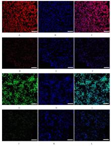
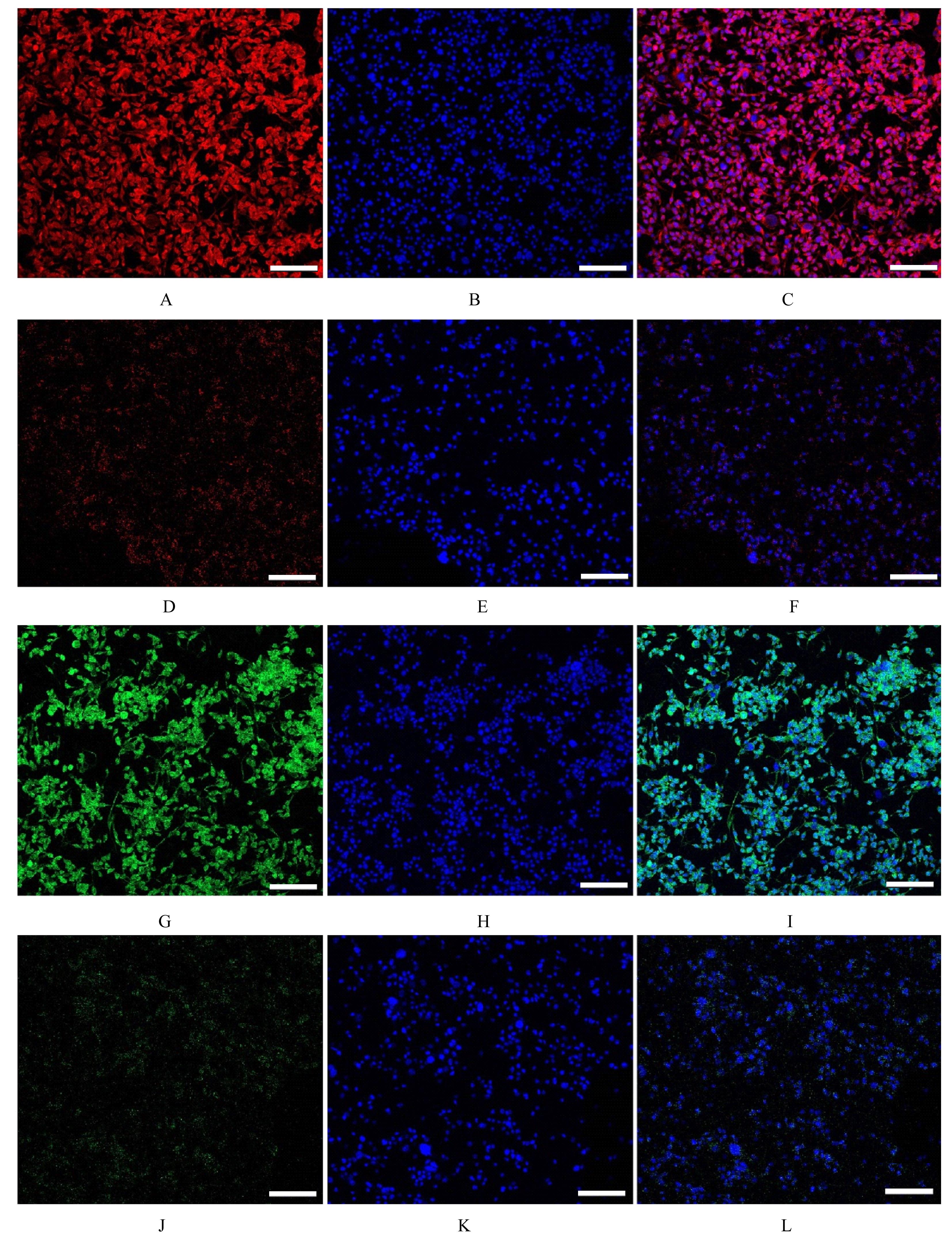
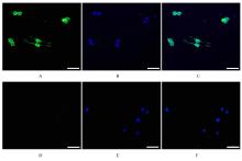
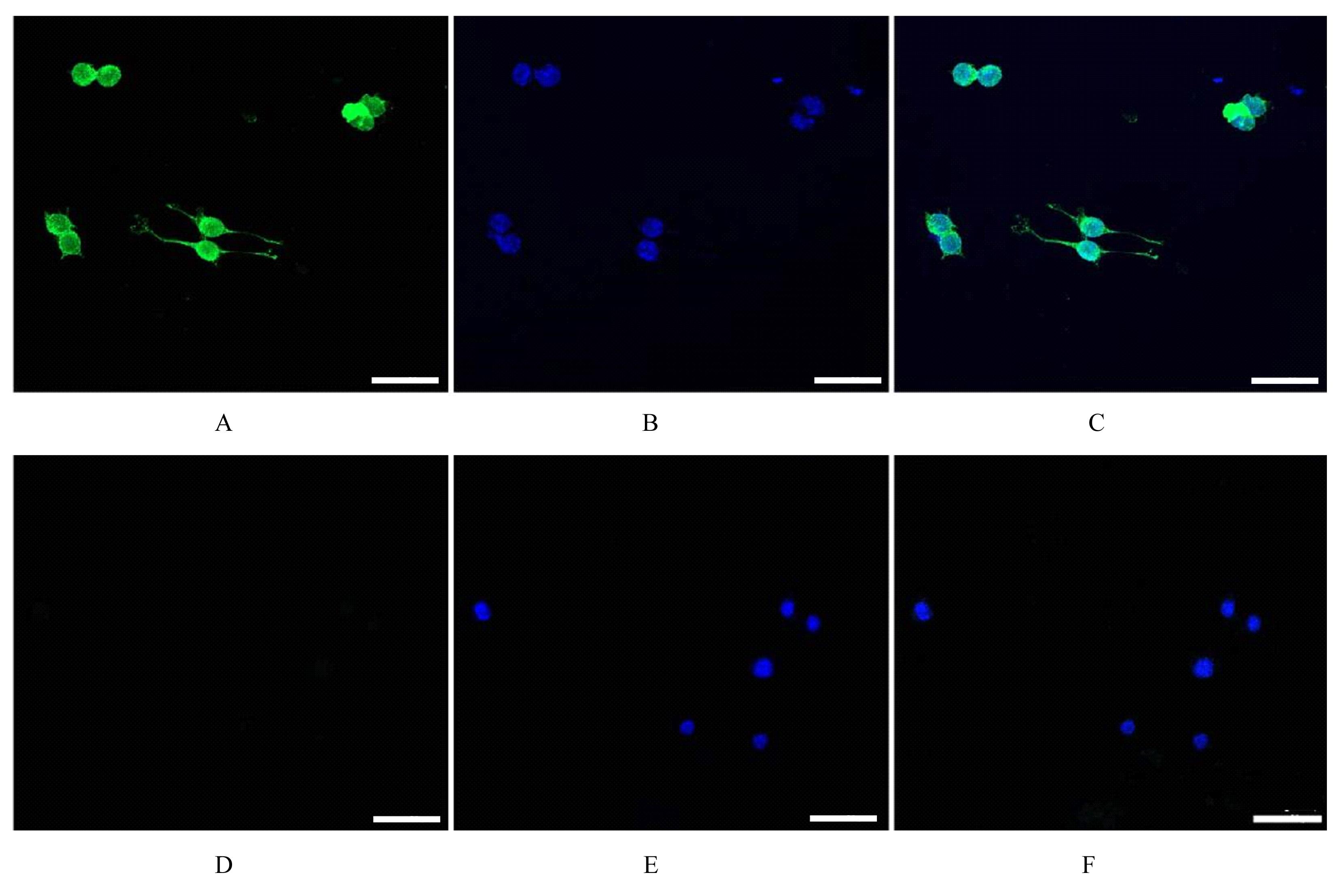
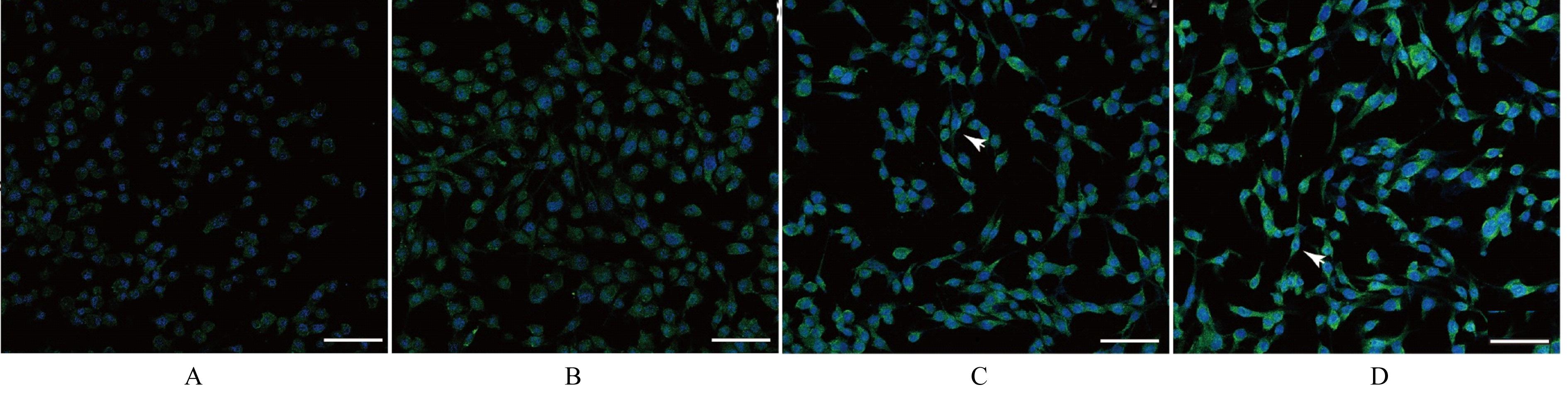
目的 观察脂多糖(LPS)诱导小鼠视网膜单独培养Müller细胞和Müller细胞与小胶质细胞共培养2种体系中的炎症反应,阐明Müller细胞与小胶质细胞的相互作用机制。 方法 培养Müller细胞QMMuC-1和小胶质细胞BV2,免疫荧光染色方法观察2种细胞形态表现。实验分为单独培养对照组[QMMuC-1细胞单独培养,采用磷酸盐(PBS)缓冲液处理]、共培养对照组(QMMuC-1细胞和BV2细胞共培养,细胞比例1∶1,采用PBS缓冲液处理)、单独培养实验组(QMMuC-1细胞单独培养,采用10 mg·L-1 LPS处理)和共培养实验组(QMMuC-1细胞和BV2细胞共培养,采用10 mg·L-1 LPS处理)。采用免疫荧光染色法观察各组细胞中胶质纤维酸性蛋白(GFAP)水平,实时荧光定量PCR(RT-qPCR)法检测各组QMMuC-1细胞中白细胞介素1β(IL-1β)、白细胞介素6(IL-6)和肿瘤坏死因子α(TNF-α)mRNA表达水平。 结果 QMMuC-1细胞中神经胶质细胞标志物谷氨酰胺合成酶(GS)和GFAP阳性,BV2细胞中小胶质细胞标志物离子钙接头蛋白分子1(Iba-1)阳性。与单独培养对照组比较,单独培养实验组QMMuC-1细胞中GFAP水平升高1.7倍(P=0.005);与共培养对照组比较,共培养实验组QMMuC-1细胞中GFAP水平升高2倍(P=0.003),细胞形态逐渐变成梭形;与单独培养实验组比较,共培养实验组QMMuC-1细胞中GFAP水平升高1.4倍(P=0.000 6),大部分细胞呈梭形。与单独培养对照组比较,单独培养实验组QMMuC-1细胞中IL-1β、IL-6和TNF-α mRNA表达水平升高,但差异无统计学意义(P>0.05);与共培养对照组比较,共培养实验组QMMuC-1细胞中IL-1β、IL-6和TNF-α mRNA表达水平升高(P<0.05);与单独培养实验组比较,共培养实验组QMMuC-1细胞中IL-1β和TNF-α mRNA表达水平升高(P<0.05)。 结论 LPS可能通过诱导小胶质细胞激活后释放炎症因子作用于Müller细胞,并加剧了Müller细胞的炎症反应。

关键词: Müller细胞, 小胶质细胞, 共培养, 脂多糖, 炎症反应

Abstract:

Objective To observe the changes of inflammation responses induced by lipopolysaccharide (LPS) in the single cultured Müller cells system and co-culture system of Müller cells and microglia of the mice, and to elucidate the interaction between the Müller cells and the microglia. Methods The Müller cells QMMuC-1 and microglia BV2 were cultured, and immunofluorescence staining was used to observe the morphology of two kinds of cells. The experiment was divided into single-culture control group [QMMuC-1 cells cultured alone, treated with phosphate buffer saline(PBS)], co-culture control group (QMMuC-1 cells and BV2 cells co-cultured with the ratio of 1∶1, treated with PBS buffer), single-culture experimental group (QMMuC-1 cells cultured alone, treated with 10 mg·L-1 LPS), and co-culture experimental group (QMMuC-1 cells and BV2 cells co-cultured, treated with 10 mg·L-1 LPS). Immunofluorescence staining was used to observe the levels of glial fibrillary acidic protein (GFAP) in the cells in various groups; real-time fluorescence quantitative PCR (RT-qPCR) method was used to detect the expression levels of interleukin-1β (IL-1β), interleukin-6 (IL-6), and tumor necrosis factor-α (TNF-α) mRNA in the QMMuC-1 cells in various groups. Results The expressions of glutamine synthetase (GS) and GFAP in the QMMuC-1 cells were positive, and the expression of ionized calcium-binding adapter molecule 1 (Iba-1) in the BV2 cells was positive. Compared with single-culture control group, the level of GFAP in the QMMuC-1 cells in single-culture experimental group was increased by 1.7 fold (P=0.005). Compared with co-culture control group, the level of GFAP in the QMMuC-1 cells in co-culture experimental group was increased by 2 fold (P=0.003),and the morphology of the cells gradually became fusiform. Compared with single-culture experimental group, the level of GFAP in the QMMuC-1 cells in co-culture experimental group was increased by 1.4 fold (P=0.0006), and most cells exhibited a fusiform shape. Compared with single-culture control group, the expression levels of IL-1β, IL-6, and TNF-α mRNA in the QMMuC-1 cells in single-culture experimental group were increased, but the expression levels had no significant differences(P>0.05). Compared with co-culture control group, the expression levels of IL-1β, IL-6, and TNF-α mRNA in the QMMuC-1 cells in co-culture experimental group were increased (P<0.05). Compared with single-culture experimental group, the expression levels of IL-1β and TNF-α mRNA in the QMMuC-1 cells in co-culture experimental group were increased (P<0.05). Conclusion LPS may induce the release of inflammatory cytokines from the activated microglia, which subsequently act on the Müller cells and exacerbate the inflammatory response in the Müller cells.

Key words: Müller cells, Microglia, Co-culture, Lipopolysaccharides, Inflammatory response

中图分类号: 

  • R774.1