吉林大学学报(医学版) ›› 2024, Vol. 50 ›› Issue (4): 1009-1015.doi: 10.13481/j.1671-587X.20240415

• 基础研究 • 上一篇    下一篇

甲磺酸阿帕替尼联合放疗对肝癌HepG2细胞的体外协同增敏作用

杨永净,柯天洋,刘士新,王雪,许德权,刘婷婷,赵玲()   

  1. 吉林省肿瘤医院放疗一科,吉林 长春 130012
  • 收稿日期:2023-07-15 出版日期:2024-07-28 发布日期:2024-08-01
  • 通讯作者: 赵玲 E-mail:2267398336@qq.com
  • 作者简介:杨永净(1975-),女,吉林省榆树市人,副主任医师,医学硕士,主要从事恶性肿瘤精准放疗方面的研究。
  • 基金资助:
    吉林省科技厅科技发展计划项目(20160101163JC);北京市希思科临床肿瘤学研究基金项目(201611234)

Synergistic sensitization of apatinib mesylate and radiotherapy on hepatocarcinoma cells invitro

Yongjing YANG,Tianyang KE,Shixin LIU,Xue WANG,Dequan XU,Tingting LIU,Ling ZHAO()   

  1. Department of Radiation Oncology,Tumor Hospital,Jilin Province,Changchun 130012,China
  • Received:2023-07-15 Online:2024-07-28 Published:2024-08-01
  • Contact: Ling ZHAO E-mail:2267398336@qq.com

摘要:

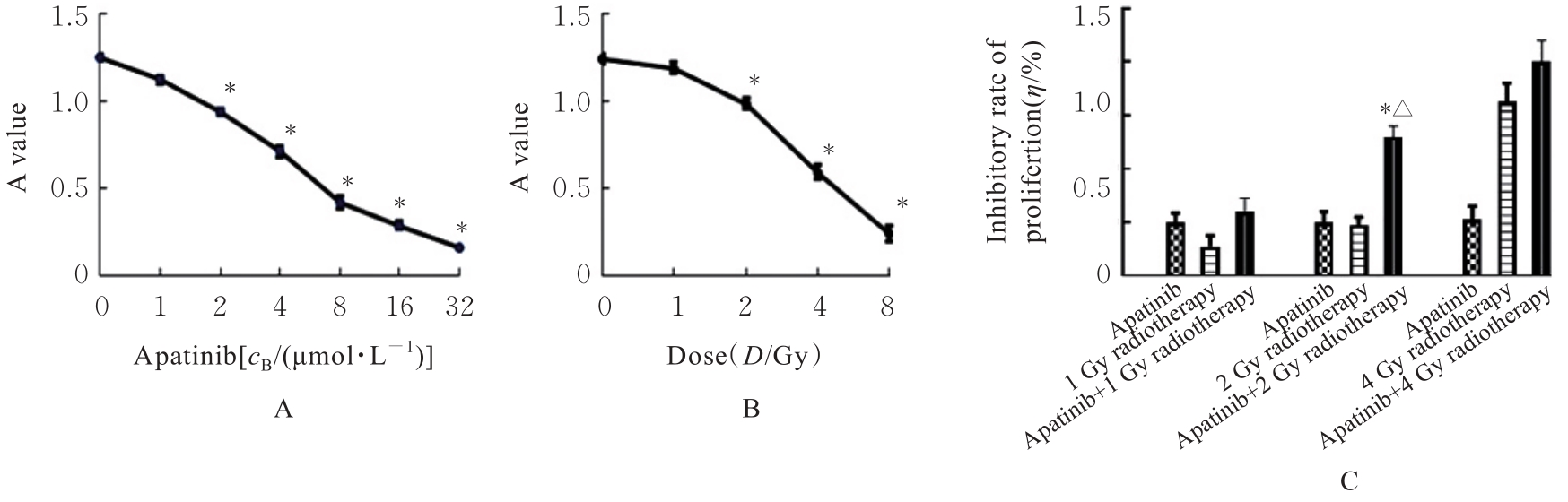
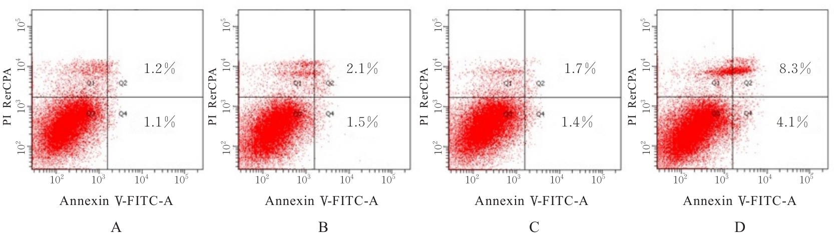
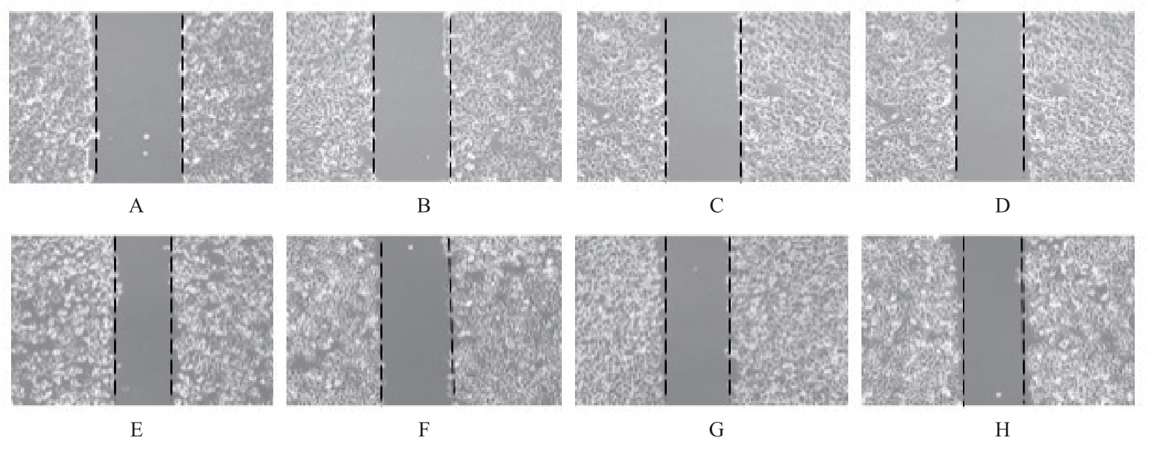
目的 探讨甲磺酸阿帕替尼(阿帕替尼)联合放射治疗(放疗)对肝癌HepG2细胞的体外协同抑制作用,阐明其相关抗肿瘤机制。 方法 体外培养肝癌HepG2细胞,以不同浓度阿帕替尼和(或)不同剂量X射线处理后,采用MTT法检测各组细胞活性,计算各组细胞增殖抑制率和阿帕替尼20%抑制浓度(IC20),并确定后续实验的X射线照射剂量。HepG2细胞分为阿帕替尼组、放疗组和阿帕替尼+放疗组(联合组),流式细胞术检测各组细胞凋亡率,细胞划痕实验检测各组细胞迁移率,酶联免疫吸附试验(ELISA)法检测各组细胞培养上清中血管内皮生长因子(VEGF)水平。 结果 MTT法检测,阿帕替尼的IC20为1.32 μmol·L-1,以此作为后续实验阿帕替尼作用浓度,确定2 Gy为后续实验X射线照射剂量。与对照组比较,阿帕替尼组和放疗组细胞凋亡率差异无统计学意义(P>0.05),联合组细胞凋亡率升高(P<0.05)。与对照组比较,阿帕替尼组、放疗组和联合组细胞迁移率降低(P<0.05);与阿帕替尼组和放疗组比较,联合组细胞迁移率降低(P<0.05)。与对照组比较,阿帕替尼组和联合组细胞培养上清中VEGF水平降低(P<0.05);与阿帕替尼和放疗组比较,联合组细胞培养上清中VEGF水平降低(P<0.05)。 结论 阿帕替尼联合放疗在体外可明显抑制肝癌HepG2细胞增殖和迁移,并诱导细胞凋亡,其作用可能与抑制细胞VEGF分泌有关。

关键词: 肝细胞癌, 甲磺酸阿帕替尼, 放射治疗, 细胞增殖, 细胞凋亡, 细胞迁移, 血管内皮细胞生长因子

Abstract:

Objective To discuss the synergistic inhibitory effect of apatinib mesylate (apatinib) combined with radiotherapy (RT) on the hepatocellular carcinoma (HepG2) cells in vitro, and to clarify its related antitumor mechanism. Methods The HepG2 cells were cultured in vitro and treated with different concentrations of apatinib and/or varying doses of X-rays. MTT method was used to detect the survival rates of the cells in various groups; the inhibitory rates of cell proliferation and the 20% inhibitory concentration (IC20) of apatinib were calculated; the X-ray irradiation dose for subsequent experiments was detected. The HepG2 cells were divided into apatinib group, RT group, and apatinib+RT group (combined group). Flow cytometry was used to detect the apoptotic rates of the cells in various groups; wound healing assay was used to detect the migration rates of the cells in various groups; ELISA method was used to detect the levels of vascular endothelial growth factor (VEGF) in the cell culture supernatant in various groups. Results The MTT results showed that the IC20 of apatinib was 1.32 μmol·L-1, and this concentration was used for subsequent experiments, and the X-ray irradiation dose for the follow-up experiments was 2 Gy. Compared with control group, the apoptotic rates of the cells in apatinib group and RT group had no significant differences (P>0.05), while the apoptotic rate of the cells in combined group was increased (P<0.05). Compared with control group, the migration rates of the cells in apatinib group, RT group, and combined group were decreased (P<0.05); compared with apatinib group and RT group, the migration rate of the cells in combined group was decreased (P<0.05). Compared with control group, the levels of VEGF in the cell culture supernatant in apatinib group and combined group were decreased (P<0.05); compared with apatinib and RT group, the level of VEGF in the cell culture supernatant in combined group was decreased (P<0.05). Conclusion Apatinib combined with radiotherapy significantly inhibits the proliferation and migration of the HepG2 cells in vitro and induces the apoptosis; its effect may be related to the inhibition of VEGF secretion by cells.

Key words: Hepatocellular carcinoma, Apatinib mesylate, Radiotherapy, Cell proliferation, Apoptosis, Cell migration, Vascular endothelial growth factor

中图分类号: 

  • R735.7