吉林大学学报(医学版) ›› 2024, Vol. 50 ›› Issue (4): 1035-1043.doi: 10.13481/j.1671-587X.20240418

• 临床研究 • 上一篇    下一篇

PLOD1在口腔鳞状细胞癌组织和细胞中的表达及其意义

郭超杰,张佳佳,曾洁,王晖予,艾尔法提·艾麦尔null,徐江()   

  1. 石河子大学第一附属医院口腔科,新疆 石河子 832000
  • 收稿日期:2023-10-11 出版日期:2024-07-28 发布日期:2024-08-01
  • 通讯作者: 徐江 E-mail:1437759520@qq.com
  • 作者简介:郭超杰(1997-),女,河南省周口市人,在读硕士研究生,主要从事颌面部肿瘤基础和临床方面的研究。
  • 基金资助:
    国家自然科学基金项目(82160572)

Expressions of PLOD1 in oral squamous cell carcinoma tissue and cells and their significances

Chaojie GUO,Jiajia ZHANG,Jie ZENG,Huiyu WANG, AIERFATI·Aimaier,Jiang XU()   

  1. Department of Stomatology,First Affiliated Hospital,Shihezi University,Shihezi 832000,China
  • Received:2023-10-11 Online:2024-07-28 Published:2024-08-01
  • Contact: Jiang XU E-mail:1437759520@qq.com

摘要:

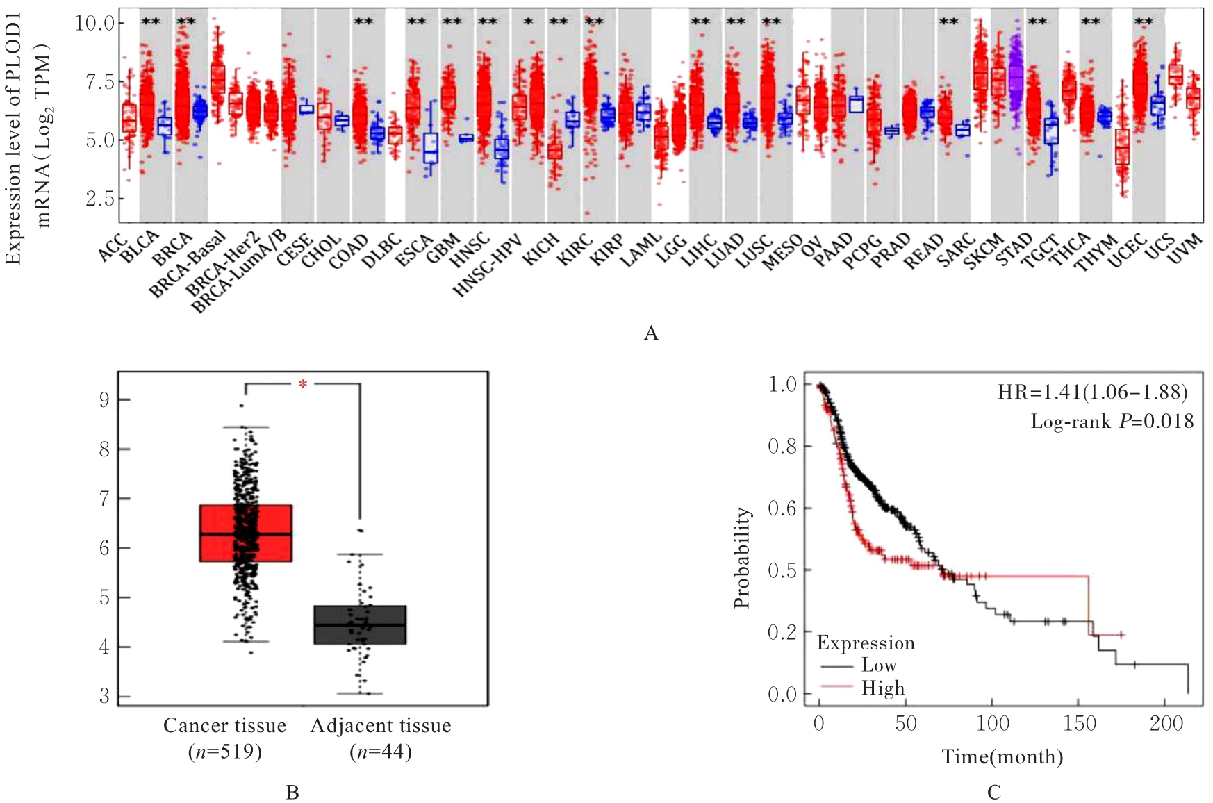
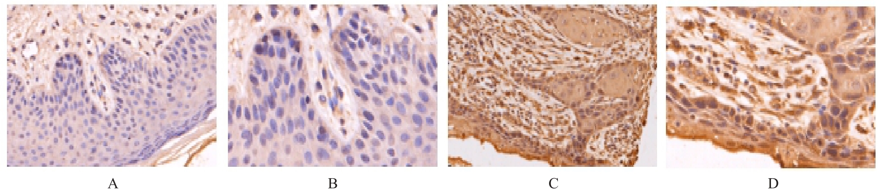
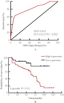
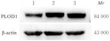
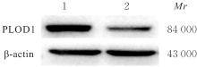
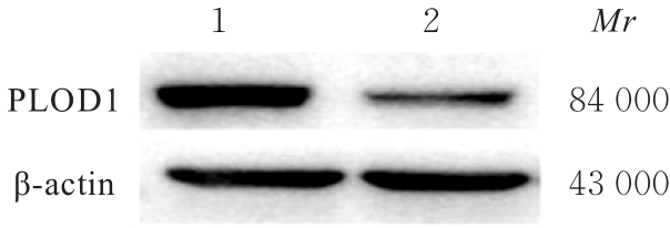
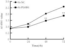
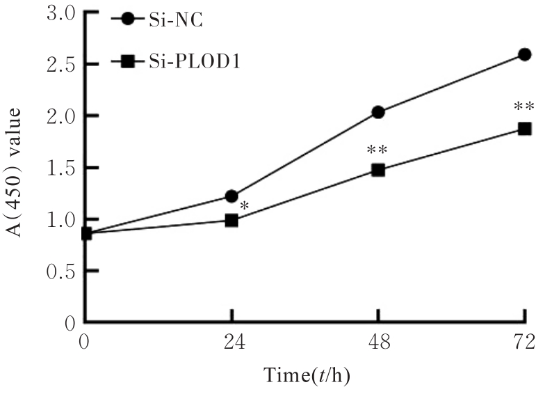
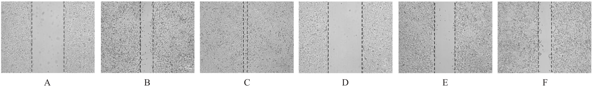
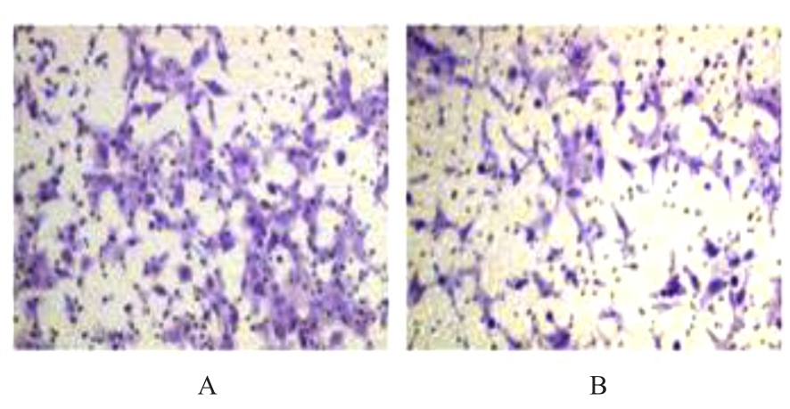
目的 探讨前胶原赖氨酸,2-酮戊二酸5-双加氧酶1(PLOD1)在口腔鳞状细胞癌(OSCC)组织和细胞中的表达及其对OSCC细胞生物学行为的影响,阐明PLOD1作为OSCC预后标志物的潜力。 方法 采用肿瘤免疫评价资源(TIMER)数据库、基因表达谱交互分析(GEPIA)数据库和Kaplan-Meier Plotter数据库分析头颈部鳞状细胞癌(HNSC)组织中PLOD1 mRMA表达水平及其与HNSC患者生存期的关联。免疫组织化学法检测110例OSCC组织和64例癌旁组织中PLOD1蛋白表达情况,分析其与OSCC患者临床病理特征和预后的关系。采用受试者工作特征(ROC)曲线下面积(AUC)评估PLOD1在OSCC诊断中的价值。实时荧光定量PCR(RT-qPCR)法和Western blotting法检测人正常口腔上皮HOK细胞和OSCC SCC15和CAL27细胞中PLOD1 mRNA和蛋白表达水平。利用脂质体法将小干扰RNA(siRNA)片段转染至SCC15细胞,分为si-NC组(转染阴性对照si-NC)和si-PLOD1组(转染si-PLOD1),采用CCK-8法、细胞划痕愈合实验和Transwell小室实验分别检测2组细胞增殖活性、划痕愈合率和侵袭细胞数。 结果 公共数据库分析,HNSC组织中PLOD1 mRNA 表达水平高于癌旁组织(P<0.05); 与 PLOD1 低表达组比较, PLOD1 高表达组HNSC患者生存期较短(HR=1.41, P=0.018)。PLOD1蛋白主要表达于 OSCC 细胞的细胞质中,OSCC组织中PLOD1 表达强度高于癌旁组织(P<0.01),并与 OSCC 患者 T 分期和 TNM 分期有关(P=0.021,P=0.004)。PLOD1 诊断 OSCC 的 AUC 为 0.811,特异度和灵敏度分别 63.64% 和90.63%。Kaplan-Meier生存分析,与PLOD1低表达组比较,PLOD1高表达组OSCC患者生存率降低(P<0.01);COX回归分析,PLOD1高表达是影响OSCC预后的独立危险因素(P=0.012)。OSCC细胞中PLOD1 mRNA和蛋白表达水平高于HOK细胞(P<0.05)。与si-NC组比较,si-PLOD1组细胞增殖活性和划痕愈合率降低(P<0.05),侵袭细胞数减少(P<0.01)。 结论 PLOD1在OSCC组织和细胞中高表达,沉默PLOD1表达可抑制OSCC细胞增殖、迁移和侵袭。PLOD1可能是OSCC新的治疗靶点和预后标志物。

关键词: 口腔鳞状细胞癌, 前胶原赖氨酸,2-酮戊二酸5-双加氧酶1, 细胞增殖, 细胞侵袭, 预后

Abstract:

Objective To discuss the expressions of procollagen-lysine, 2-oxoglutarate 5-dioxygenase 1 (PLOD1) in oral squamous cell carcinoma (OSCC) tissue and OSCC cells and its effect on the biological behavior of the OSCC cells, and to clarify the potential of PLOD1 as a prognostic biomarker for OSCC. Methods The expression level of PLOD1 mRNA in head and neck squamous cell carcinoma (HNSC) tissue and its correlation with the survival of the HNSC patients were analyzed by Tumor Immune Estimation Resource (TIMER), Gene Expression Profiling Interactive Analysis (GEPIA), and Kaplan-Meier Plotter databases. Immunohistochemistry method was used to detect the expression level of PLOD1 protein in 110 OSCC tissue and 64 adjacent tissue,and its association with clinicopathological characteristics and prognosis of the OSCC patients were analyzed; the diagnostic value of PLOD1 in OSCC was detected by area under the curve (AUC) of the receiver operating characteristic (ROC). The expression levels of PLOD1 mRNA and protein in the human normal oral epithelial HOK cells and OSCC SCC15 and CAL27 cells were detected by real-time fluorescence quantitative PCR (RT-qPCR) and Western blotting methods. The small interfering RNA (siRNA) fragments were transfected into the SCC15 cells by liposome method and the cells were dividing into si-NC group (transfected with negative control si-NC) and si-PLOD1 group (transfected with si-PLOD1); the proliferation activities, scratch healing rates, and numbers of invasion cells in two groups were detected by CCK-8 method, wound healing assay, and Transwell chamber assay, respectively. Results The public database analysis results showed that the expression level of PLOD1 mRNA in HNSC tissue was higher than that in adjacent tissue(P<0.05); compared with low expression of PLOD1 group, the survival period of the HNSC patients in high expression of PLOD1 group was shorter (HR=1.41, P=0.018). The PLOD1 protein was mainly expressed in cytoplasm of the OSCC cells, and the expression intensity of PLOD1 in OSCC tissue was higher than that in adjacent tissue (P<0.01), and it was related to T stage and TNM stage of the OSCC patients (P=0.021, P=0.004). The AUC of PLOD1 for diagnosing OSCC was 0.811, and the specificity was 63.64% and the sensitivity was 90.63%. The Kaplan-Meier survival analysis results showed that compared with low expression of PLOD1 group, the survival rate of the OSCC patients in high expression of PLOD1 group was decreased (P<0.01).The COX regression analysis results showed that high expression of PLOD1 was an independent risk factor for the prognosis of OSCC (P=0.012). The expression levels of PLOD1 mRNA and protein in the OSCC cells were higher than those in HOK cells (P<0.05). Compared with si-NC group, the proliferation activity and scratch healing rate of the cells in si-PLOD1 group were decreased (P<0.05), and the number of invasion cells was decreased(P<0.01). Conclusion PLOD1 is highly expressed in the OSCC tissue and cells, and silencing of PLOD1 expression can inhibit the proliferation, migration, and invasion of the OSCC cells. PLOD1 may be a new therapeutic target and prognostic biomarker for OSCC.

Key words: Oral squamous cell carcinoma, Procollagen-lysine, 2-oxoglutarate 5-dioxygenase 1, Cell proliferation, Cell invasion, Prognosis

中图分类号: 

  • R739.8